Learn about our comprehensive antibody validation methods to ensure monospecificity. Antibody Validation>>

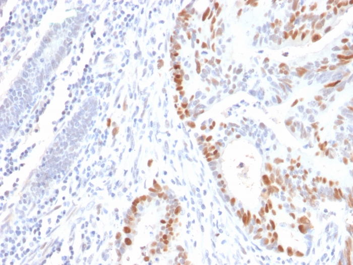
Formalin-fixed, paraffin-embedded human Colon Carcinoma stained with p53 Mouse Recombinant Monoclonal Antibody (rBP53-12).

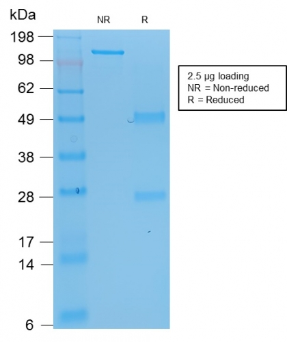
SDS-PAGE Analysis of Purified p53 Mouse Recombinant Monoclonal Antibody (rBP53-12). Confirmation of Integrity and Purity of Antibody.

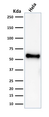
Western Blot Analysis of HeLa cell lysate using p53 Mouse Recombinant Monoclonal Antibody (rBP53-12).

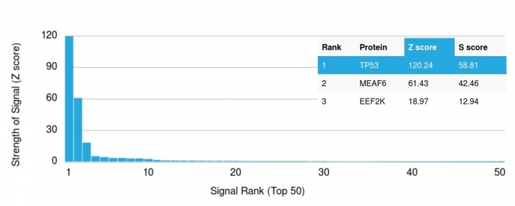
Analysis of Protein Array containing more than 19,000 full-length human proteins using p53 Recombinant Mouse Monoclonal Antibody (rBP53-12). Z- and S- Score: The Z-score represents the strength of a signal that a monoclonal antibody (MAb) (in combination with a fluorescently-tagged anti-IgG secondary antibody) produces when binding to a particular protein on the HuProtTM array. Z-scores are described in units of standard deviations (SD's) above the mean value of all signals generated on that array. If targets on HuProtTM are arranged in descending order of the Z-score, the S-score is the difference (also in units of SD's) between the Z-score. S-score therefore represents the relative target specificity of a MAb to its intended target. A MAb is considered to specific to its intended target, if the MAb has an S-score of at least 2.5. For example, if a MAb binds to protein X with a Z-score of 43 and to protein Y with a Z-score of 14, then the S-score for the binding of that MAb to protein X is equal to 29.
This MAb reacts with an N-terminal epitope (aa 16-25) of both wild type and mutated p53. Mutation and/or allelic loss of p53 is one of the causes of a variety of mesenchymal and epithelial tumors. If it occurs in the germ line, such tumors run in families. In most transformed and tumor cells the concentration of p53 is increased 51000 fold over the minute concentrations (1000 molecules cell) in normal cells, principally due to the increased half-life (4 h) compared to that of the wild-type (20 min). p53 Localizes in the nucleus, but is detectable at the plasma membrane during mitosis and when certain mutations modulate cytoplasmic/nuclear distribution. Mutations arise with an average frequency of 70% but incidence varies from zero in carcinoid lung tumors to 97% in primary melanomas. High concentrations of p53 protein are transiently expressed in human epidermis and superficial dermal fibroblasts following mild ultraviolet irradiation. Positive nuclear staining with p53 antibody has been reported to be a negative prognostic factor in breast carcinoma, lung carcinoma, colorectal, and urothelial carcinoma. Anti-p53 positivity has also been used to differentiate uterine serous carcinoma from endometrioid carcinoma as well as to detect intratubular germ cell neoplasia.
There are no reviews yet.