Immunohistochemistry (IHC) is a powerful technique for visualizing specific proteins in tissue sections, providing valuable insights for research and diagnostics. This protocol outlines the steps for tissue rehydration, antigen retrieval, and the IHC process, which often involves utilizing antibodies.
Materials
Tissue sections on slides.
Slide Brite
Alcohol Series: Ethanol (95% and 70%) for rehydration.
Antigen Retrieval Solution: Choose the appropriate method (e.g., heat-induced epitope retrieval or enzyme digestion).
Blocking Solution: Prepare a blocking solution to prevent non-specific binding (e.g., serum-based or protein-based).
Primary Antibodies: Obtain specific primary antibodies for your target proteins.
Secondary Antibodies: Choose suitable secondary antibodies conjugated with enzymes or fluorophores.
Detection System: Depending on your choice, this can include enzyme-linked detection (DAB) or fluorescence detection.
Counterstain: Hematoxylin for nuclear counterstaining.
Mounting Medium: To preserve the slides.
Microscope: For visualization.
*Make sure to use personal protective equipment, such as gloves and lab coats.
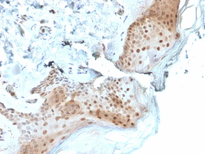
Formalin-fixed, paraffin-embedded human Skin stained with Stratifin Mouse Monoclonal Antibody (CPTC-SFN-2).
Formalin-fixed, paraffin-embedded human Testis stained with 14-3-3E Mouse Monoclonal Antibody (CPTC-YWHAE-1).
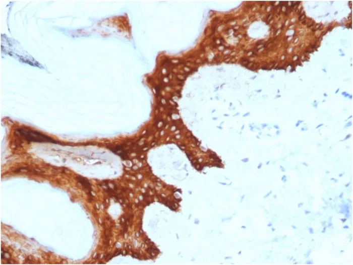
Formalin-fixed, paraffin-embedded human Skin stained with 8-oxoguanine Mouse Monoclonal Antibody (CPTC-OGG1-1).
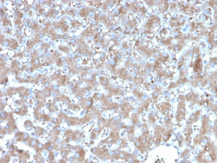
Formalin-fixed, paraffin-embedded human hepatocellular carcinoma stained with TNFAIP3 Mouse Monoclonal Antibody (TNFAIP3/2813).
Label the tissue slides according to the protocol. Arrange the slides well in slide racks for easy handling.
Place the slide racks in an oven preheated to 60 °C. Bake the slides for 15 minutes or until the paraffin on the tissue loosens and forms droplets.
Deparaffinize and rehydrate tissue sections by immersing slides through the following series of solutions.
Perform 3 changes of Slide Brite, each for 5 minutes.
Perform 2 changes of 100% ethanol, each for an appropriate duration.
Perform 2 changes of 95% ethanol, each for 5 minutes.
Immerse the slides in 80% ethanol for 5 minutes.
Submerge the slides in 50% ethanol for 5 minutes.
Perform 2 changes of distilled water, each for 5 minutes.
Perform 2 changes of 3% hydrogen peroxide, each for 5 minutes.
Antigen Retrieval
Antigen retrieval can be achieved using BioCare’s Decloacker, which involves boiling the sections at 95 °C for 45 minutes using Tris-EDTA (10 mM Tris with 1 mM EDTA, pH 9.0) OR citrate buffer (10 mM citrate pH 6.0).
Immunohistochemistry
Utilize a hydrophobic marker to establish protective barriers above and below the tissue sections. Exercise caution during this process, as marking too close to the tissue may potentially lead to tissue drying. Therefore, it is advisable to refrain from marking too close to the tissue during this step.
Wash the tissue section twice, 3 times each in PBS.
Incubate the sections with the blocking solution to minimize non-specific binding. Incubation time varies but is typically 30 minutes to 1 hour.
Apply the specific primary antibody to the sections. Dilute the antibody according to the manufacturer’s recommendations. Incubate at the recommended temperature and time (usually overnight at 4 °C or 30 minutes to 2 hours at room temperature).
Wash the sections twice with PBS, 3–5 minutes each, to remove unbound primary antibody.
Apply the appropriate secondary antibody conjugated with an enzyme or fluorophore. Incubate for the recommended time and temperature. Incubate for 30 minutes with secondary antibody, goat anti-mouse / goat anti-rabbit-HRP polymer.
Wash the slides to remove unbound secondary antibody.
Apply the detection system as per your choice. (e.g., DAB for enzyme-linked detection or fluorescent dye for fluorescence detection). For DAB staining, incubate the slides in the DAB solution for 3–5 minutes.
DAB preparation: Mix 950 μL of DAB substrate and 500 μL of DAB chromogen (ScyTek Laboratories) well and use immediately.
Counterstain the nuclei with hematoxylin 3–5 times.
Wash sections with PBS for 3–5 minutes. The final wash should be with distilled water.
Dehydrate through an alcohol series (70%, 95%, and 100% ethanol) and xylene. Mount the slides with a mounting medium and coverslip.
Observe cells under a microscope.
Analyze the stained tissue sections under a microscope and assess the cellular protein localization and expression levels in tissues, which is vital for research and diagnostics.
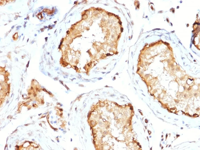
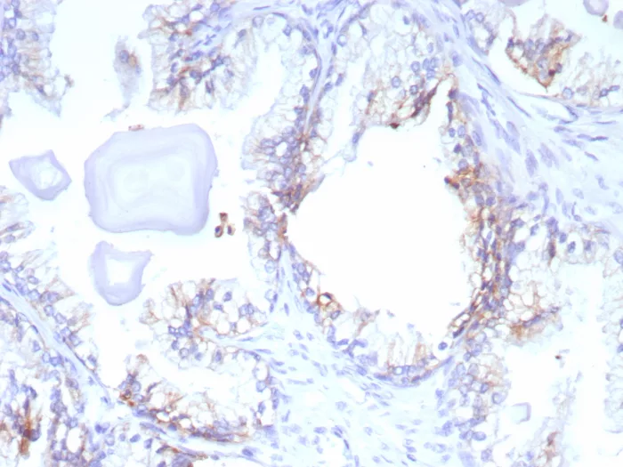
Formalin-fixed, paraffin-embedded human prostate cancer stained with ABCC4 Mouse Monoclonal Antibody (ABCC4/9179). HIER: Tris/EDTA, pH9.0, 45min. 2°C: HRP-polymer, 30min. DAB, 5min.
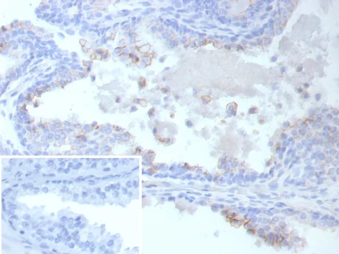
Formalin-fixed, paraffin-embedded human prostate cancer stained with ABCC4 Mouse Monoclonal Antibody (ABCC4/9019). Inset: PBS instead of primary antibody; secondary only negative control.
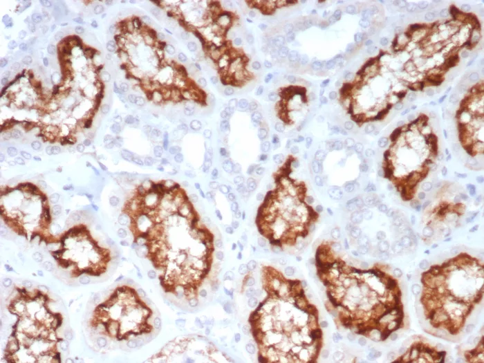
IHC analysis of formalin-fixed, paraffin-embedded human kidney. Stained using ACE2/7203 at 2ug/ml in PBS for 30min RT. HIER: Tris/EDTA, pH9.0, 45min. 2°C: HRP-polymer, 30min. DAB, 5min.
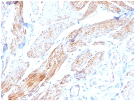
Formalin-fixed, paraffin-embedded human bladder stained with Aciculin Mouse Monoclonal Antibody (PGM5/3552) at 2ug/ml. HIER: Tris/EDTA, pH9.0, 45min. 2 °: HRP-polymer, 30min. DAB, 5min.