Free Shipping in the U.S. for orders over $1000. Shop Now>>

SDS-PAGE Analysis of Purified p27 Recombinant Rabbit Monoclonal Antibody (KIP1/1355R). Confirmation of Purity and Integrity of Antibody.

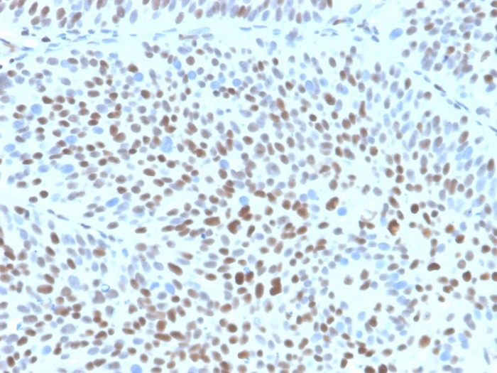
IHC analysis of formalin-fixed, paraffin-embedded human bladder. Strong nuclear staining using KIP1/1355R at 2ug/ml in PBS for 30min RT. HIER: Tris/EDTA, pH9.0, 45min. 2°C: HRP-polymer, 30min. DAB, 5min.
This MAb recognizes a 27kDa protein, identified as the p27Kip1, a cell cycle regulatory mitotic inhibitor. It is highly specific and shows no cross-reaction with other related mitotic inhibitors. In Western blotting of cell lysates from 7 human breast cancer cell lines (ZR75-1, ZR75-30, MCF-7, MDAMB453, T47D, CAL51, 734B), the antibody labels a single band corresponding to p27Kip1. It functions as a negative regulator of G1 progression and has been proposed to function as a possible mediator of TGF- � � induced G1 arrest. p27Kip1 is a candidate tumor suppressor gene. Reportedly, low p27 expression has been associated with unfavorable prognosis in renal cell carcinoma, colon carcinoma, breast carcinomas, non-small-cell lung carcinoma, hepatocellular carcinoma, multiple myeloma, and lymph node metastases in papillary carcinoma of the thyroid, as well as a more aggressive phenotype in carcinoma of the cervix.
There are no reviews yet.