| Catalog Number: | 3005-RBM5 |
|---|---|
| Gene: | |
| Reactivity: | |
| Host: |
Trial Size Available
Carrier-Free Available

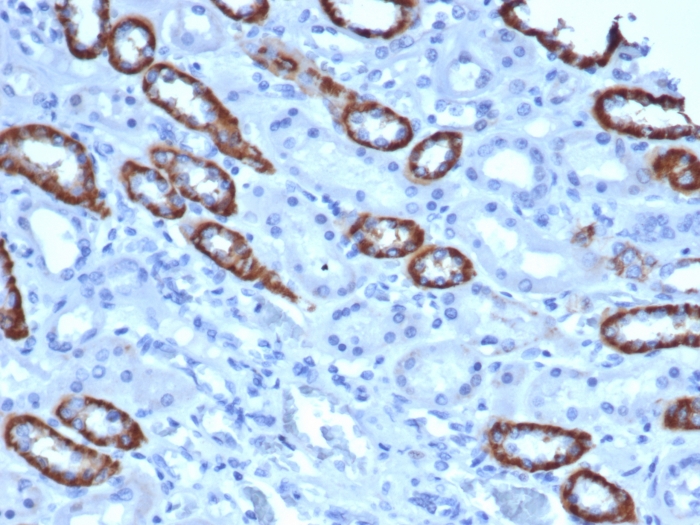
Formalin-fixed, paraffin-embedded human Skin Basal Cell Carcinoma stained with Histone H1 Rabbit Recombinant Monoclonal Antibody (AE-4).

Formalin-fixed, paraffin-embedded human Colon Carcinoma stained with Histone H1 Rabbit Recombinant Monoclonal Antibody (AE-4).

Formalin-fixed, paraffin-embedded human Breast Carcinoma stained with Histone H1 Rabbit Recombinant Monoclonal Antibody (AE-4).
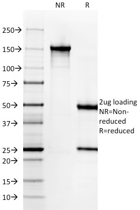
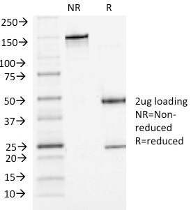
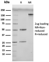
SDS-PAGE Analysis Purified Histone H1 Rabbit Recombinant Monoclonal Antibody (AE-4). Confirmation of Purity and Integrity of Antibody.
| Document Name | |
|---|---|
| Datasheet | Download Here |
| Material Safety Data Sheet | Download Here |
| Applications | Tested Dilutions | Protocol | Note |
|---|---|---|---|
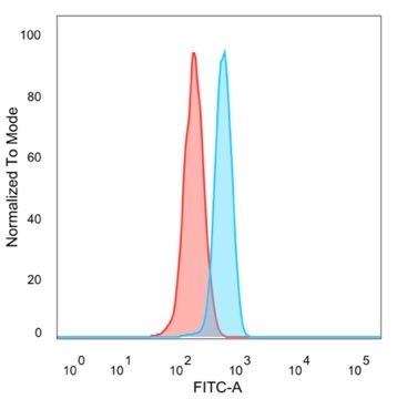
| Flow Cytometry (Flow) | 1-2ug/million cells | Flow Cytometry Protocol | |
| Immunofluorescence (IF) | 1-3ug/ml | ||
| Immunohistochemistry (IHC) | 1-2ug/ml | IHC Protocol | 30 min at RT. Staining of formalin-fixed tissues requires heating tissue sections in 10mM Tris with 1mM EDTA, pH 9.0, for 45 min at 95°C followed by cooling at RT for 20 minutes |
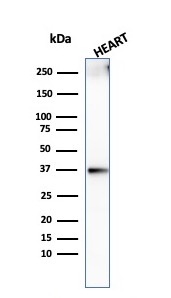
| Western Blot (WB) | 2-4ug/ml | Western Blot Protocol |
Please note that this antibody is a recombinant Rabbit version of original anti-histone H1 antibody (Clone AE-4). Because the variable heavy (VH) and variable light (VL) domains are the same, recombinant antibody has the same exact reactivity as the original AE-4 MAb. There are several advantages of producing a recombinant version of a monoclonal antibody. For example, a recombinant antibody is a pure preparation of active immunoglobulin with no contaminating non-functional intact Ig or free light/heavy chains. Secondly, antibody can always be produced, even if hybridoma line is lost. Moreover, it adds the flexibility of converting the antibody to any species, isotype or format. Eukaryotic histones are basic and water-soluble nuclear proteins that form hetero-octameric nucleosome particles by wrapping 146 base pairs of DNA in a left-handed super-helical turn sequentially to form chromosomal fiber. Two molecules of each of the four core histones (H2A, H2B, H3, and H4) form the octamer; formed of two H2A-H2B dimers and two H3-H4 dimers, forming two nearly symmetrical halves by tertiary structure. Over 80% of nucleosomes contain the linker Histone H1, derived from an intronless gene that interacts with linker DNA between nucleosomes and mediates compaction into higher order chromatin. This antibody is extensively used as a pan-nuclear marker.
Should a NeoBiotechnologies antibody not meet the experimental expectations outlined on our website or datasheet, please contact our Technical Support. We unconditionally guarantee our product.
Our team of scientists are ready to help you with expert recommendations or technical assistance related to applications or product use.
Information
© 2026 NeoBiotechnologies. All Rights Reserved.
Monday-Friday: 8:00am – 8:00pm Pacific Time