Learn about our comprehensive antibody validation methods to ensure monospecificity. Antibody Validation>>

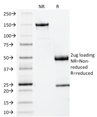
SDS-PAGE Analysis of Purified Beta-2-Microglobulin Mouse Monoclonal Antibody (BBM.1). Confirmation of Integrity and Purity of Antibody.

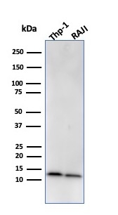
Western Blot Analysis of human THP-1 and Raji cell lysate using Beta-2-Microglobulin Mouse Monoclonal Antibody (BBM.1)
Recognizes a protein of 12kDa, identified as microglobulin. Major histocompatibility complex (MHC) class 1 molecules bind to antigens for presentation on the surface of cells. The proteasome is responsible for producing these antigens from the components of foreign pathogens. MHC class 1 molecules consist of an alpha heavy chain that contains three subdomains (alpha1, alpha2, alpha3) and a non-covalent associating light chain, known as beta-2-Microglobulin. Beta-2-Microglobulin associates with the alpha3 subdomain of the alpha heavy chain and forms an immunoglobulin domain-like structure that mediates proper folding and expression of MHC class 1 molecules. The alpha1 and alpha2 domains of the alpha heavy chain form the peptide antigen-binding cleft. Mutations in the beta-2-Microglobulin gene can enhance the progression of malignant melanoma phenotypes.
There are no reviews yet.