Learn about our comprehensive antibody validation methods to ensure monospecificity. Antibody Validation>>

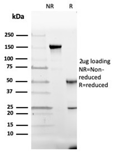
SDS-PAGE Analysis of Purified GC Mouse Monoclonal Antibody (VDBP/4482). Confirmation of Purity and Integrity of Antibody.

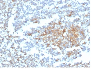
IHC analysis of formalin-fixed, paraffin-embedded human tonsil. Staining using VDBP/4482 at 2ug/ml in PBS for 30min RT. HIER: Tris/EDTA, pH9.0, 45min. 2°C: HRP-polymer, 30min. DAB, 5min.

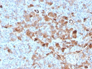
IHC analysis of formalin-fixed, paraffin-embedded human pancreas. Staining using VDBP/4482 at 2ug/ml in PBS for 30min RT.

Analysis of Protein Array containing more than 19,000 full-length human proteins using GC Vitamin D Binding Protein Mouse Monoclonal Antibody (VDBP/4482). Z- and S- Score: The Z-score represents the strength of a signal that a monoclonal antibody (MAb) (in combination with a fluorescently-tagged anti-IgG secondary antibody) produces when binding to a particular protein on the HuProtTM array. Z-scores are described in units of standard deviations (SD's) above the mean value of all signals generated on that array. If targets on HuProtTM are arranged in descending order of the Z-score, the S-score is the difference (also in units of SD's) between the Z-score. S-score therefore represents the relative target specificity of a MAb to its intended target. A MAb is considered to specific to its intended target, if the MAb has an S-score of at least 2.5. For example, if a MAb binds to protein X with a Z-score of 43 and to protein Y with a Z-score of 14, then the S-score for the binding of that MAb to protein X is equal to 29.
Vitamin D-binding protein (DBP) is a multi-functional serum protein that binds to the plasma membranes of numerous cell types and mediates a variety of cellular functions. The locus of the DBP protein (also known as group-specific component protein or GC) is located at human chromosome 4q13.3. DBP functions in organ-specific transportation of vitamin D and its metabolites to the various target organs of the vitamin D endocrine system. In addition, DBP has immunomodulatory properties and is able to bind to the surface of leukocytes. DBP binds to the plasma membrane through a chondroitin sulfate proteoglycan. DBP serves as a co-chemotactic factor for C5a to enhance the chemotactic activity of C5a. DBP can also bind to globular Actin with high affinity and is involved in the clearance of Actin from the blood. DBP plays an important role in osteoclast differentiation. The diverse cellular functions of DBP require its cell surface binding ability to mediate different biological processes.
There are no reviews yet.