2 Union Square, Union City, CA 94587
contact@NeoBiotechnologies.com
510-376-5603

Home >> Antibodies >> CD43 (T-cell Marker)

CD43 (T-cell Marker)

Mouse Monoclonal Antibody [Clone SPM503]

Catalog #

Size

20 ug
100 ug
100 ug

Price (USD)

219.00
499.00
499.00

Formulation

Purified Ab with BSA and Azide at 200ug/ml
Purified Ab WITHOUT BSA and Azide at 1.0mg/ml

Catalog No

Size

20 ug
100 ug
100 ug

Formulation

Purified Ab with BSA and Azide at 200ug/ml
Purified Ab WITHOUT BSA and Azide at 1.0mg/ml

Price (USD)

219.00
499.00
499.00
img

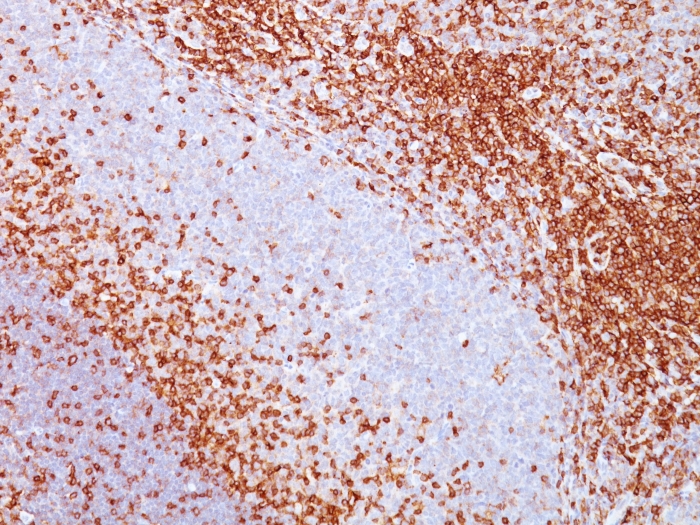
Formalin-fixed, paraffin-embedded human tonsil stained with CD43 Mouse Monoclonal Antibody (SPM503).
img

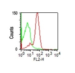
Flow cytometric analysis of human PBMCs using CD43 Mouse Monoclonal Antibody (SPM504); isotype control (green).

img

Immunofluorescence Analysis of K562 cells labeling CD43 with CD43 Mouse Monoclonal Antibody (SPM503) followed by goat anti-mouse IgG-CF488 (green). Nuclear counterstain is NucSpotŪ Live 650.

img

Western Blot Analysis of K562 cell lysate using CD43 Mouse Monoclonal Antibody (SPM503).

Product Details

Synonyms

Galactoglycoprotein, GALGP, GPL115, Leukocyte sialoglycoprotein, Leukosialin, LSN, Sialophorin, SPN

Positive Control

PBMCs or K562 cells (FC). K562 cell lysate (WB). Paracortex in a tonsil or a reactive lymph node (IHC).

Known Applications & Suggested Dilutions

Flow Cyt, IF, Western Blot, IHC-P         More Details

Flow Cytometry (1-2ug/million cells)
Immunofluorescence (1-2ug/ml)
Western Blot (1-2ug/ml)
Immunohistochemistry (Formalin-fixed) (1-2ug/ml for 30 minutes at RT)(Staining of formalin-fixed tissues requires heating tissue sections in 10mM Tris with 1mM EDTA, pH 9.0, for 45 min at 95 °C followed by cooling at RT for 20 minutes)Optimal dilution for a specific application should be determined.

Immunogen

Myeloblastic KG1 cells

Cellular Localization

cell surface

Species Reactivity

Human.

Host / Ig Isotype

Mouse / IgG1, kappa

Mol. Weight of Antigen

95, 115, or 135kDa

Specificity & Comments

It recognizes a cell surface glycoprotein of 95/115/135kDa (depending upon the extent of glycosylation), identified as CD43. 70-90% of T-cell lymphomas and from 22-37% of B-cell lymphomas express CD43. No reactivity has been observed with reactive B-cells. So, a B-lineage population that co-expresses CD43 is highly likely to be a malignant lymphoma, especially a low-grade lymphoma, rather than a reactive B-cell population. When CD43 antibody is used in combination with anti-CD20, effective immunophenotyping of the lymphomas in formalin-fixed tissues can be obtained. Co-staining of a lymphoid infiltrate with anti-CD20 and anti-CD43 argues against a reactive process and favors a diagnosis of lymphoma.

Related Products


200ug/ml of Ab purified from Bioreactor Concentrate by Protein A/G. Prepared in 10mM PBS with 0.05% BSA & 0.05% azide. Also available WITHOUT BSA & azide at 1.0mg/ml.

Key References

  1. Stross WP, et. al. Journal of Clinical Pathology, 1989, 42(9):953-61.

Bioinformatics

  •  Entrez Gene ID: 6693
  •  Gene Symbol: SPN
  •  SwissProt: P16150
  •  Chromosome Location: 16p11.2
  •  Unigene: 632188