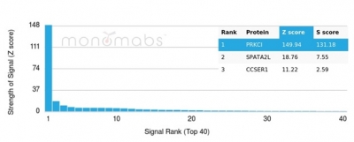
Learn about our comprehensive antibody validation methods to ensure monospecificity.  Antibody Validation>>

Protein Kinase C iota / lambda / PRKCI Antibody [PRKCI/4912]

In Stock
HuProt Validated
Catalog Number Formulation Size Price
5584-MSM2-P0
Purified Ab with BSA and Azide at 200ug/ml
20ug
$229.00
5584-MSM2-P1
Purified Ab with BSA and Azide at 200ug/ml
100ug
$519.00
5584-MSM2-P1ABX
Purified Ab WITHOUT BSA and Azide at 1.0mg/ml
100ug
$519.00
Flat Rate Domestic: $60 | Orders outside the US - Contact Us for Order Information | Ships next business day

Applications & Dilutions

Applications Tested Dillution Protocol Note
Immunohistochemistry (IHC)
1-2ug/ml
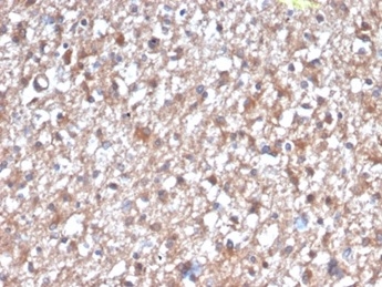
30 min at RT. Staining of formalin-fixed tissues requires heating tissue sections in 10mM Tris with 1mM EDTA, pH 9.0, for 45 min at 95°C followed by cooling at RT for 20 minutes

Summary

Members of the protein kinase C (PKC) family play a key regulatory role in a variety of cellular functions, including cell growth and differentiation, gene expression, hormone secretion and membrane function. PKCs were originally identified as serine/threonine protein kinases whose activity was dependent on calcium and phospholipids. Diacylglycerols (DAG) and tumor promoting phorbol esters bind to and activate PKC. PKCs can be subdivided into at least two major classes, including conventional (c) PKC isoforms (伪, 尾I, 尾II and 纬) and novel (n) PKC isoforms ( �L, , �梅, , , l/i, m and n). Patterns of expression for each PKC isoform differ among tissues and PKC family members exhibit clear differences in their cofactor dependencies. For instance, the kinase activities of PKC �L and are independent of Ca2+. On the other hand, most of the other PKC members possess phorbol ester-binding activities and kinase activities.

Product Properties & Targets

Antibody Type
Host
Mouse
Applications
Species Reactivity
Isotype / Light Chain
IgG2b / Kappa
Cellular Localization
Cytoplasm, Endosome, Membrane, Nucleus
Gene Name
Positive Control
Brain, Liver or placenta.
Immunogen
Recombinant fragment (around aa100-300) of human PRKCI protein (exact sequence is proprietary)
Alternate Names
Protein kinase C iota type, Atypical protein kinase C-lambda/iota, nPKC-iota, aPKC lambda/iota; Atypical protein kinase C lambda/iota; Atypical protein kinase C-lambda/iota; nPKC-iota; nPKCiota; PKC lambda/iota; PKCI; PKCiota; PKClambda; PRKC iota; PRKC lambda/iota; PRKCI; Protein kinase C iota type

Database Links

Entrez Gene ID
SwissProt

Additional Information

Clone
PRKCI/4912
Chromosome Location
3q26.2
Mol. Weight of Antigen
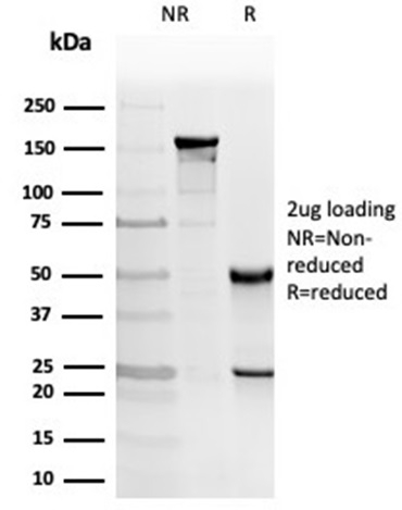
68kDa

Functions

  • Calcium- and diacylglycerol-independent serine/ threonine-protein kinase that plays a general protective role against apoptotic stimuli, is involved in NF-kappa-B activation, cell survival, differentiation and polarity, and contributes to the regulation of microtubule dynamics in the early secretory pathway. Is necessary for BCR-ABL oncogene-mediated resistance to apoptotic drug in leukemia cells, protecting leukemia cells against drug-induced apoptosis. In cultured neurons, prevents amyloid beta protein-induced apoptosis by interrupting cell death process at a very early step. In glioblastoma cells, may function downstream of phosphatidylinositol 3-kinase (PI(3)K) and PDPK1 in the promotion of cell survival by phosphorylating and inhibiting the pro-apoptotic factor BAD. Can form a protein complex in non-small cell lung cancer (NSCLC) cells with PARD6A and ECT2 and regulate ECT2 oncogenic activity by phosphorylation, which in turn promotes transformed growth and invasion. In response to nerve growth factor (NGF), acts downstream of SRC to phosphorylate and activate IRAK1, allowing the subsequent activation of NF-kappa-B and neuronal cell survival. Functions in the organization of the apical domain in epithelial cells by phosphorylating EZR. This step is crucial for activation and normal distribution of EZR at the early stages of intestinal epithelial cell differentiation. Forms a protein complex with LLGL1 and PARD6B independently of PARD3 to regulate epithelial cell polarity. Plays a role in microtubule dynamics in the early secretory pathway through interaction with RAB2A and GAPDH and recruitment to vesicular tubular clusters (VTCs). In human coronary artery endothelial cells (HCAEC), is activated by saturated fatty acids and mediates lipid-induced apoptosis. Involved in early synaptic long term potentiation phase in CA1 hippocampal cells and short term memory formation (By similarity).

Key References

  • Bicaku, E., et al. 2005. Cyclin-dependent kinase activating kinase/Cdk7 co-localizes with PKC-i in human glioma cells. Tissue Cell 37: 53-58.

PubMed Links

Storage & Stability

Antibody with azide - store at 2 to 8 °C. Antibody without azide - store at -20 to -80 °C. Antibody is stable for 24 months. Non-hazardous. No MSDS required.

Limitations

This antibody is available for research use only and is not approved for use in diagnosis.

Supplied as

200ug/ml of Ab purified from Bioreactor Concentrate by Protein A/G. Prepared in 10mM PBS with 0.05% BSA & 0.05% azide. Also available WITHOUT BSA & azide at 1.0mg/ml.

Warranty

There are no warranties, expressed or implied, which extend beyond this description. Company is not liable for any personal injury or economic loss resulting from this product.

Reviews

There are no reviews yet.

Be the first to review “Protein Kinase C iota / lambda / PRKCI Antibody”

Your email address will not be published. Required fields are marked *

PARTNERSHIP OPPORTUNITIES

We hold Exclusive rights to 10,000 recombinant and hybridoma antibody products, available for Licensing or Collaboration.

LETS TALK