Learn about our comprehensive antibody validation methods to ensure monospecificity. Antibody Validation>>

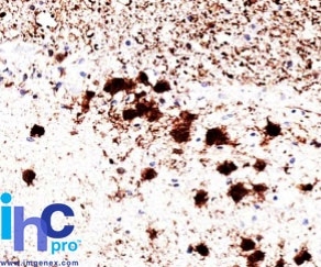
Formalin-fixed, paraffin-embedded human brain stained with Pgp9.5 / UchL1 Mouse Monoclonal Antibody (31A3).

Western blot of UchL1 (PGP9.5) in 1) human, 2) Mouse and 3) rat brain lysate using Pgp9.5 / UchL1 Mouse Monoclonal Antibody (31A3).

Western Blot Analysis of human brain tissue lysate using Pgp9.5 / UchL1 Mouse Monoclonal Antibody (31A3).
This MAb reacts with a protein of 20-30kDa, identified as PGP9.5, also known as ubiquitin carboxyl-terminal hydrolase-1 (UchL1). Initially, PGP9.5 expression in normal tissues was reported in neurons and neuroendocrine cells but later it was found in distal renal tubular epithelium, spermatogonia, Leydig cells, oocytes, melanocytes, prostatic secretory epithelium, ejaculatory duct cells, epididymis, mammary epithelial cells, Merkel cells, and dermal fibroblasts. Furthermore, immunostaining for PGP9.5 has been shown in a wide variety of mesenchymal neoplasms as well. A mutation in PGP9.5 gene is believed to cause a form of Parkinson's disease.
There are no reviews yet.