Learn about our comprehensive antibody validation methods to ensure monospecificity.  Antibody Validation>>

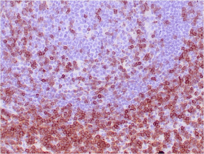
CD7 (T-Cell Leukemia Marker) Antibody [CD7/3737]

In Stock
Catalog Number Formulation Size Price
924-MSM7-P0
Purified Ab with BSA and Azide at 200ug/ml
20ug
$229.00
924-MSM7-P1
Purified Ab with BSA and Azide at 200ug/ml
100ug
$519.00
924-MSM7-P1ABX
Purified Ab WITHOUT BSA and Azide at 1.0mg/ml
100ug
$519.00
Flat Rate Domestic: $60 | Orders outside the US - Contact Us for Order Information | Ships next business day

Applications & Dilutions

Applications Tested Dillution Protocol Note
Immunohistochemistry (IHC)
1-2ug/ml
30 min at RT. Staining of formalin-fixed tissues requires heating tissue sections in 10mM Tris with 1mM EDTA, pH 9.0, for 45 min at 95°C followed by cooling at RT for 20 minutes
Western Blot (WB)
2-4ug/ml

Summary

Recognizes a protein of 40kDa, identified as CD7 (also known as gp40, Leu9). CD7 is a member of the immunoglobulin gene superfamily. Its N-terminal amino acids 1-107 are highly homologous to Ig kappa-L chains whereas the carboxyl-terminal region of the extracellular domain is proline-rich and has been postulated to form a stalk from which the Ig domain projects. CD7 is expressed on the majority of immature and mature T-lymphocytes, and T cell leukemia. It is also found on natural killer cells, a small subpopulation of normal B cells and on malignant B cells. Cross-linking surface CD7 positively modulates T cell and NK cell activity as measured by calcium fluxes, expression of adhesion molecules, cytokine secretion and proliferation. CD7 associates directly with phosphoinositol 3'-kinase. CD7 ligation induces production of D-3 phosphoinositides and tyrosine phosphorylation.

Product Properties & Targets

Antibody Type
Host
Mouse
Species Reactivity
Isotype / Light Chain
IgG1 / Kappa
Cellular Localization
Membrane
Gene Name
CD7
Positive Control
HePG-2 or Jurkat cells. Human lymph node or tonsil.
Immunogen
Recombinant fragment (around aa 72-175) of human CD7 protein (exact sequence is proprietary)
Alternate Names
T-cell antigen CD7, GP40, T-cell leukemia antigen, T-cell surface antigen Leu-9, TP41, GP40; Leu9; p41; T-cell leukemia antigen; T-cell surface antigen Leu-9; Tp40; TP41

Database Links

Entrez Gene ID
924
SwissProt

Additional Information

Clone
CD7/3737
Chromosome Location
17q25.3
Mol. Weight of Antigen
40kDa

Functions

  • Not yet known.

Key References

  • Saxena A, et al. Biologic and clinical significance of CD7 expression in acute myeloid leukemia. Am J Hematol. 1998;58(4):278-84.

Storage & Stability

Antibody with azide - store at 2 to 8 °C. Antibody without azide - store at -20 to -80 °C. Antibody is stable for 24 months. Non-hazardous. No MSDS required.

Limitations

This antibody is available for research use only and is not approved for use in diagnosis.

Supplied as

200ug/ml of Ab purified from Bioreactor Concentrate by Protein A/G. Prepared in 10mM PBS with0.05% BSA & 0.05% azide. Also available WITHOUT BSA & azide at 1.0mg/ml.

Warranty

There are no warranties, expressed or implied, which extend beyond this description. Company is not liable for any personal injury or economic loss resulting from this product.

Reviews

There are no reviews yet.

Be the first to review “CD7 (T-Cell Leukemia Marker) Antibody”

Your email address will not be published. Required fields are marked *

PARTNERSHIP OPPORTUNITIES

We hold Exclusive rights to 10,000 recombinant and hybridoma antibody products, available for Licensing or Collaboration.

LETS TALK