Learn about our comprehensive antibody validation methods to ensure monospecificity. Antibody Validation>>

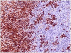
Formalin-fixed, paraffin-embedded human tonsil stained with CD138Recombinant Rabbit Monoclonal Antibody (SDC1/4378R).

Formalin-fixed, paraffin-embedded human tonsil stained with CD138 Recombinant Rabbit Monoclonal Antibody (SDC1/4378R).
CD138, also designated syndecan-1, is a member of the syndecan family of four transmembrane spanning proteins capable of binding to heparan sulfate and chondroitin sulfate molecules. The syndecans main functions are to control cell growth and differentiation as well as to maintain cell adhesion and cell migration. Under normal conditions CD138 ispredominantly expressed on mature plasma cells and early preB-cells, while other haematolymphoid cells are negative. Various types of epithelial cells are also CD138 positive. Squamous epithelial cells show strong membranous and some cytoplasmic staining, except for the mature superficial squamous epithelial cells which are unstained. Mature mesenchymal and neural tissues are not stained. Among haematolymphoid neoplasms, CD138 is expressed in practically all cases of plasma cell malignancies. Among non-haematolymphoid neoplasms the expression of CD138 is found in various types of carcinoma. In the following, the large majority of cases is CD138 positive: skin squamous cell carcinoma and basal cell carcinoma, colorectal adenocarcinoma, cholangiocarcinoma, transitional cell carcinoma, endometrial adenocarcinoma, ductal and lobular breast carcinoma, hepatocellular carcinoma, renal cell carcinoma, and lung adenocarcinoma.CD138 is a very sensitive and specific marker for identification of plasma cells and plasma cell differentiation within haematolymphoid tissues in benign and neoplastic conditions.
There are no reviews yet.