Learn about our comprehensive antibody validation methods to ensure monospecificity. Antibody Validation>>

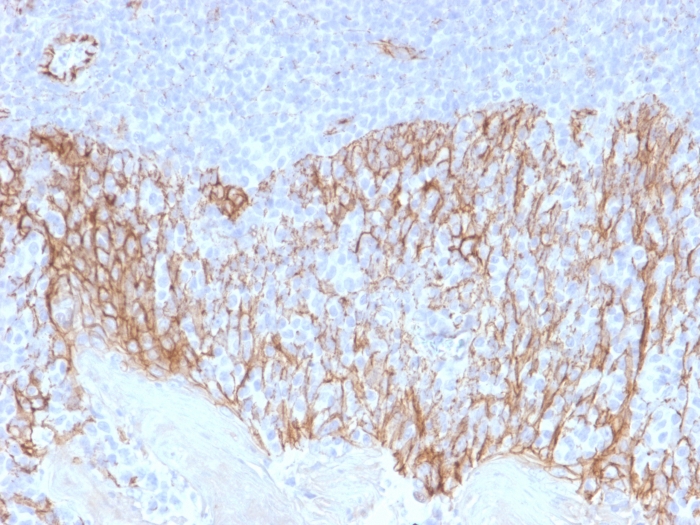
Formalin-fixed, paraffin-embedded human Tonsil stained with Beta-Catenin Mouse Monoclonal Antibody (CTNNB1/2099).

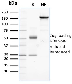
SDS-PAGE Analysis of Purified Beta-Catenin Mouse Monoclonal Antibody (CTNNB1/2099). Confirmation of Integrity and Purity of Antibody.

Analysis of Protein Array containing more than 19,000 full-length human proteins using Catenin, beta (CTNNB1) Mouse Monoclonal Antibody (CTNBB1/2099). Z- and S- Score: The Z-score represents the strength of a signal that a monoclonal antibody (Monoclonal Antibody) (in combination with a fluorescently-tagged anti-IgG secondary antibody) produces when binding to a particular protein on the HuProtTM array. Z-scores are described in units of standard deviations (SD's) above the mean value of all signals generated on that array. If targets on HuProtTM are arranged in descending order of the Z-score, the S-score is the difference (also in units of SD's) between the Z-score. S-score therefore represents the relative target specificity of a Monoclonal Antibody to its intended target. A Monoclonal Antibody is considered to specific to its intended target, if the Monoclonal Antibody has an S-score of at least 2.5. For example, if a Monoclonal Antibody binds to protein X with a Z-score of 43 and to protein Y with a Z-score of 14, then the S-score for the binding of that Monoclonal Antibody to protein X is equal to 29.
Beta-catenin associates with the cytoplasmic portion of E-cadherin, which is necessary for the function of E-cadherin as an adhesion molecule. In normal tissues, beta-catenin is localized to the membrane of epithelial cells, consistent with its role in the cell adhesion complex. In breast ductal neoplasia, beta-catenin is usually localized in cellular membranes. However, in lobular neoplasia, a marked redistribution of beta-catenin throughout the cytoplasm results in a diffuse cytoplasmic pattern. Immuno-staining of beta-catenin and E-cadherin is helps in the accurate identification of ductal and lobular neoplasms, including a distinction between low-grade ductal carcinoma in situ (DCIS) and lobular carcinoma. Additionally, some rectal and gastric adenocarcinomas demonstrate diffuse cytoplasmic beta-catenin staining and a lack of membranous staining, mimicking the staining pattern observed with lobular breast carcinomas.
There are no reviews yet.