Learn about our comprehensive antibody validation methods to ensure monospecificity. Antibody Validation>>

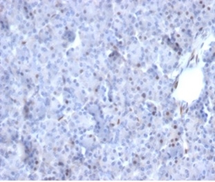
Formalin-fixed, paraffin-embedded human pancreas stained with AKT1 Mouse Monoclonal Antibody (AKT1/2784). HIER: Tris/EDTA, pH9.0, 45min. 2 °: HRP-polymer, 30min. DAB, 5min.

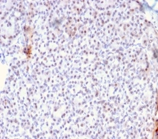
Formalin-fixed, paraffin-embedded human pancreas stained with AKT1 Mouse Monoclonal Antibody (AKT1/2784).

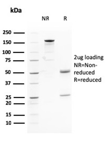
SDS-PAGE Analysis Purified AKT1 Mouse Monoclonal Antibody (AKT1/2784). Confirmation of Purity and Integrity of Antibody.

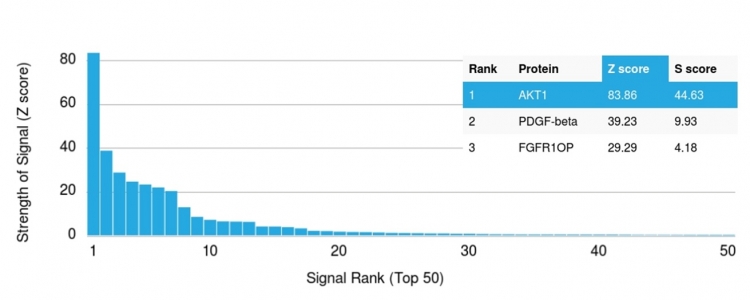
Analysis of Protein Array containing more than 19,000 full-length human proteins using AKT1-Monospecific Mouse Monoclonal Antibody (AKT1/2784). Z- and S- Score: The Z-score represents the strength of a signal that a monoclonal antibody (MAb) (in combination with a fluorescently-tagged anti-IgG secondary antibody) produces when binding to a particular protein on the HuProtTM array. Z-scores are described in units of standard deviations (SDâs) above the mean value of all signals generated on that array. If targets on HuProtTM are arranged in descending order of the Z-score, the S-score is the difference (also in units of SDâs) between the Z-score. S-score therefore represents the relative target specificity of a MAb to its intended target. A MAb is considered to specific to its intended target, if the MAb has an S-score of at least 2.5. For example, if a MAb binds to protein X with a Z-score of 43 and to protein Y with a Z-score of 14, then the S-score for the binding of that MAb to protein X is equal to 29.
Recognizes a protein of 62kDa, which is identified as AKT1. The serine/threonine kinase Akt family contains several members, including Akt1 (also designated PKB or RacPK), Akt2 (also designated PKB尾 or RacPK-尾) and Akt 3 (also designated PKB纬 or thymoma viral proto-oncogene 3), which exhibit sequence homology with the protein kinase A and C families and are encoded by the c-Akt proto-oncogene. All members of the Akt family have a Pleckstrin homology domain. Akt1 and Akt2 are activated by PDGF stimulation. This activation is dependent on PDGFR-尾 tyrosine residues 740 and 751, which bind the subunit of the phosphatidylinositol 3-kinase (PI 3-kinase) complex. Activation of Akt1 by insulin or insulin-growth factor-1 (IGF-1) results in phosphorylation of both Thr 308 and Ser 473. Akt proteins become phosphorylated and activated in insulin/IGF-1-stimulated cells by an upstream kinase(s), and the activation of Akt1 and Akt2 is inhibited by the PI kinase inhibitor wortmannin.
There are no reviews yet.