Learn about our comprehensive antibody validation methods to ensure monospecificity. Antibody Validation>>

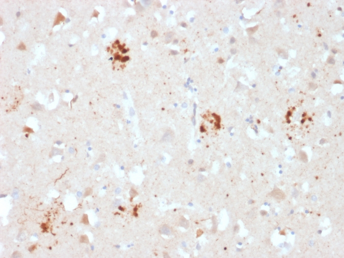
Formalin-fixed, paraffin-embedded human Brain stained with Ubiquitin Mouse Monoclonal Antibody (UBB/2122).

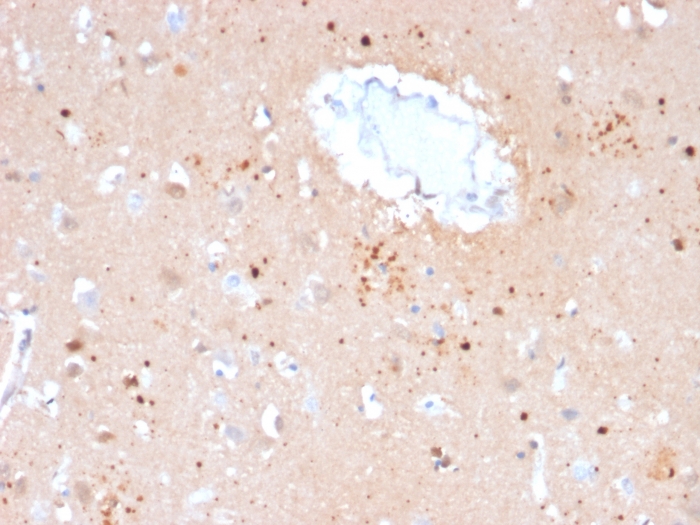
Formalin-fixed, paraffin-embedded human Brain stained with Ubiquitin Mouse Monoclonal Antibody (UBB/2122).

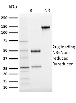
SDS-PAGE Analysis Purified Ubiquitin Mouse Monoclonal Antibody (UBB/2122). Confirmation of purity and integrity.

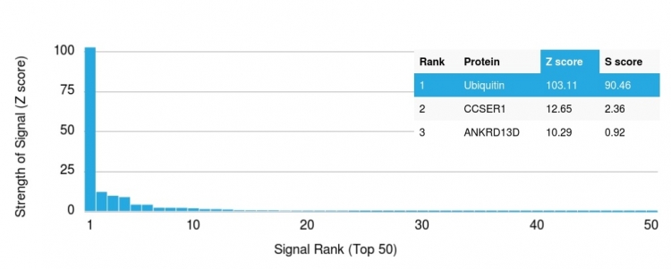
Analysis of Protein Array containing more than 19,000 full-length human proteins using Ubiquitin Mouse Monoclonal Antibody (UBB/2122) Z- and S- Score: The Z-score represents the strength of a signal that a monoclonal antibody (MAb) (in combination with a fluorescently-tagged anti-IgG secondary antibody) produces when binding to a particular protein on the HuProtTM array. Z-scores are described in units of standard deviations (SD's) above the mean value of all signals generated on that array. If targets on HuProtTM are arranged in descending order of the Z-score, the S-score is the difference (also in units of SD's) between the Z-score. S-score therefore represents the relative target specificity of a MAb to its intended target. A MAb is considered to specific to its intended target, if the MAb has an S-score of at least 2.5. For example, if a MAb binds to protein X with a Z-score of 43 and to protein Y with a Z-score of 14, then the S-score for the binding of that MAb to protein X is equal to 29.
Ubiquitin is a highly conserved and plays an essential role in the ubiquitin-proteasome pathway. In ubiquitination process, it is first activated by forming a thiol-ester complex with the activation component E1, which is then transferred to ubiquitin-carrier protein E2, followed by to ubiquitin ligase E3 for final delivery to epsilon-NH2 of the target protein lysine residue. IkB, p53, cdc25A, Bcl-2 etc. are shown as targets of ubiquitin-proteasome process as part of regulation of cell cycle progression, differentiation, cell stress response, and apoptosis. Moreover, ubiquitin have been reported to bind covalently with pathological inclusions which are resistant to degradation e.g. neurofibrillary tangles/paired helical filaments in Alzheimer’s disease, Lewy bodies seen in Parkinson’s disease, and Pick bodies found in Pick’s disease etc.
There are no reviews yet.