2 Union Square, Union City, CA 94587
contact@NeoBiotechnologies.com
510-376-5603

Home >> Antibodies >> Neuronal-Nuclei (NeuN) (Neuronal Marker)

Neuronal-Nuclei (NeuN) (Neuronal Marker)

Recombinant Rabbit Monoclonal Antibody [Clone NeuN/288R]

Catalog #

Size

20 ug
100 ug
100 ug

Price (USD)

219.00
499.00
499.00

Formulation

Purified Ab with BSA and Azide at 200ug/ml
Purified Ab WITHOUT BSA and Azide at 1.0mg/ml

Catalog No

Size

20 ug
100 ug
100 ug

Formulation

Purified Ab with BSA and Azide at 200ug/ml
Purified Ab WITHOUT BSA and Azide at 1.0mg/ml

Price (USD)

219.00
499.00
499.00
img

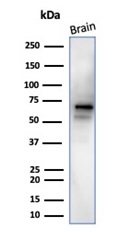
Western Blot Analysis of human brain tissue lysate using NeuN Recombinant Rabbit Monoclonal Antibody (NeuN/288R).
img

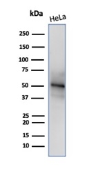
Western blot analysis of HeLa cell lysate using NeuN Recombinant Rabbit Monoclonal Antibody (NeuN/288R).

img

Formalin-fixed, paraffin-embedded human cerebrum stained with NeuN Recombinant Rabbit Monoclonal Antibody (NeuN/288R). HIER: Tris/EDTA, pH9.0, 45min. 2°: HRP-polymer, 30min. DAB, 5min.

img

Formalin-fixed, paraffin-embedded human cerebellum stained with NeuN Recombinant Rabbit Monoclonal Antibody (NeuN/288R). Inset: PBS instead of primary antibody, secondary negative control.

Product Details

Synonyms

FLJ56884; FLJ58356; fox1 homolog C; Fox3; FOX3NeuN; hexaribonucleotide binding protein 3 (HRNBP3); neuronal nuclei; Rbfox3; RNA binding protein, fox 1 homolog (C. elegans) 3

Positive Control

Human brain. HeLa cells.

Known Applications & Suggested Dilutions

Western Blot, IHC-P         More Details

Western Blot (1-2ug/ml)
Immunohistochemistry (Formalin-fixed) (1-2ug/ml for 30 minutes at RT)
(Staining of formalin-fixed tissues requires heating tissue sections in 10mM Tris with 1mM EDTA, pH 9.0, for 45 min at 95 °C followed by cooling at RT for 20 minutes)
Optimal dilution for a specific application should be determined.

Immunogen

Recombinant full-length human NeuN protein

Cellular Localization

Nucleus. Cytoplasm.

Species Reactivity

Human and mouse.

Host / Ig Isotype

Rabbit / IgG, kappa

Mol. Weight of Antigen

48kDa

Specificity & Comments

NeuN antibody specifically recognizes the DNA-binding, neuron-specific protein NeuN, which is present in most CNS and PNS neuronal cell types of all vertebrates tested. NeuN protein distributions are apparently restricted to neuronal nuclei and some proximal neuronal processes in both fetal and adult brain although, some neurons fail to be recognized by NeuN at all ages: INL retinal cells, Cajal-Retzius cells, Purkinje cells, inferior olivary and dentate nucleus neurons, and sympathetic ganglion cells are examples. Immunohistochemically detectable NeuN protein first appears at developmental timepoints that correspond with the withdrawal of the neuron from the cell cycle and/or with the initiation of terminal differentiation of the neuro. Immunoreactivity appears around E9.5 in the mouse neural tube and is extensive throughout the developing nervous system by E12.5. Strong nuclear staining suggests a nuclear regulatory protein function; however, no evidence currently exists as to whether the NeuN protein antigen has a function in the distal cytoplasm or whether it is merely synthesized there before being transported back into the nucleus. No difference between protein isolated from purified nuclei and whole brain extract on immunoblots has been found.

Related Products


200ug/ml of Ab purified from Bioreactor Concentrate by Protein A/G. Prepared in 10mM PBS with 0.05% BSA & 0.05% azide. Also available WITHOUT BSA & azide at 1.0mg/ml.

Key References

  1. Edgar MA, et al. Arch Pathol Lab Med.2008; 132:500-9.
  2. Ulbright TM, et al. Mod Pathol.2010; 23:972-80.

Bioinformatics

  •  Entrez Gene ID: 146713
  •  Gene Symbol: RBFOX3
  •  SwissProt: A6NFN3
  •  Chromosome Location: 17q25.3
  •  Unigene: 135229