Free Shipping in the U.S. for orders over $1000. Shop Now>>

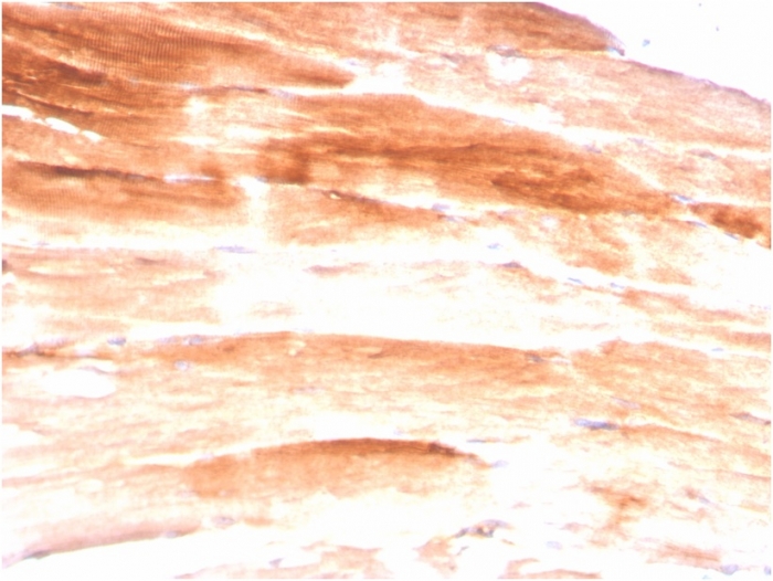
Formalin-fixed, paraffin-embedded human Skeletal Muscle stained with Dystrophin Monospecific Mouse Monoclonal Antibody (DMD/3245).

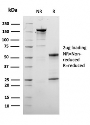
SDS-PAGE Analysis of Purified Dystrophin Monospecific Mouse Monoclonal Antibody (DMD/3245). Confirmation of Purity and Integrity of Antibody.

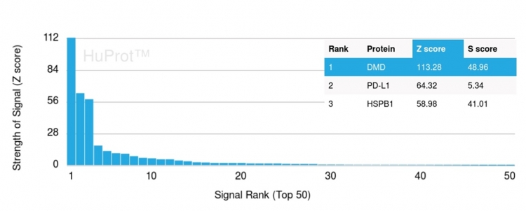
Analysis of Protein Array containing more than 19,000 full-length human proteins using Dystrophin Monospecific Mouse Monoclonal Antibody (DMD/3245). Z- and S- Score: The Z-score represents the strength of a signal that a monoclonal antibody (Monoclonal Antibody) (in combination with a fluorescently-tagged anti-IgG secondary antibody) produces when binding to a particular protein on the HuProtTM array. Z-scores are described in units of standard deviations (SD's) above the mean value of all signals generated on that array. If targets on HuProtTM are arranged in descending order of the Z-score, the S-score is the difference (also in units of SD's) between the Z-score. S-score therefore represents the relative target specificity of a Monoclonal Antibody to its intended target. A Monoclonal Antibody is considered to specific to its intended target, if the Monoclonal Antibody has an S-score of at least 2.5. For example, if a Monoclonal Antibody binds to protein X with a Z-score of 43 and to protein Y with a Z-score of 14, then the S-score for the binding of that Monoclonal Antibody to protein X is equal to 29.
Dystrophin-glycoprotein complex (DGC) connects the F-Actin cytoskeleton on the inner surface of muscle fibers to the surrounding extracellular matrix, through the cell membrane interface. A deficiency in this protein contributes to Duchenne (DMD) and Becker (BMD) muscular dystrophies. The human dystrophin gene measures 2.4 megabases, has more than 80 exons, produces a 14 kb mRNA and contains at least 8 independent tissue-specific promoters and 2 poly A sites. The dystrophin mRNA can undergo differential splicing and produce a range of transcripts that encode a large set of proteins. Dystrophin represents approximately 0.002% of total striated muscle protein and localizes to triadic junctions in skeletal muscle, where it is thought to influence calcium ion homeostasis and force transmission.
There are no reviews yet.