2 Union Square, Union City, CA 94587
contact@NeoBiotechnologies.com
510-376-5603

Home >> Antibodies >> CD7 (T-Cell Leukemia Marker)

CD7 (T-Cell Leukemia Marker)

Mouse Monoclonal Antibody [Clone CD7/3737]

Catalog #

Size

20 ug
100 ug
100 ug

Price (USD)

219.00
499.00
499.00

Formulation

Purified Ab with BSA and Azide at 200ug/ml
Purified Ab WITHOUT BSA and Azide at 1.0mg/ml

Catalog No

Size

20 ug
100 ug
100 ug

Formulation

Purified Ab with BSA and Azide at 200ug/ml
Purified Ab WITHOUT BSA and Azide at 1.0mg/ml

Price (USD)

219.00
499.00
499.00
img

Formalin-fixed, paraffin-embedded human lymph node stained with CD7 Mouse Monoclonal Antibody (CD7/3737).
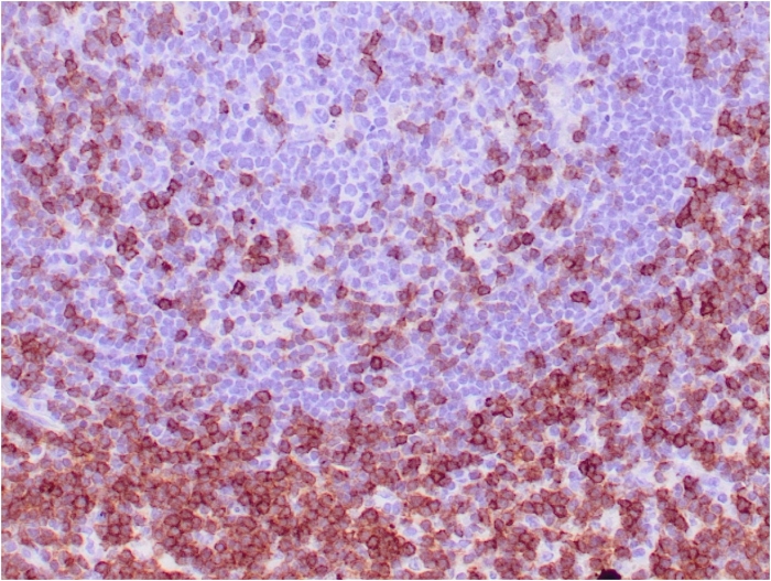
img

Formalin-fixed, paraffin-embedded human tonsil stained with CD7 Mouse Monoclonal Antibody (CD7/3737).

img

Formalin-fixed, paraffin-embedded human lymph node stained with CD7 Mouse Monoclonal Antibody (CD7/3737).

img

Western blot analysis of HePG-2 cell lysate using CD7 Mouse Monoclonal Antibody (CD7/3737).

img

SDS-PAGE Analysis Purified CD7 Mouse Monoclonal Antibody (CD7/3737). Confirmation of Purity and Integrity of Antibody.

Product Details

Synonyms

GP40; Leu9; p41; T-cell leukemia antigen; T-cell surface antigen Leu-9; Tp40; TP41

Positive Control

HePG-2 or Jurkat cells. Human lymph node or tonsil.

Known Applications & Suggested Dilutions

Western Blot, IHC-P         More Details

Western Blot (1-2ug/ml)
Immunohistochemistry (Formalin-fixed) (1-2ug/ml for 30 minutes at RT)
(Staining of formalin-fixed tissues requires heating tissue sections in 10mM Tris with 1mM EDTA, pH 9.0, for 45 min at 95 °C followed by cooling at RT for 20 minutes)
Optimal dilution for a specific application should be determined.

Immunogen

Recombinant fragment (around aa 72-175) of human CD7 protein (exact sequence is proprietary)

Cellular Localization

Cell Surface

Species Reactivity

Human.

Host / Ig Isotype

Mouse / IgG1, kappa

Mol. Weight of Antigen

40kDa

Specificity & Comments

Recognizes a protein of 40kDa, identified as CD7 (also known as gp40, Leu9). CD7 is a member of the immunoglobulin gene superfamily. Its N-terminal amino acids 1-107 are highly homologous to Ig kappa-L chains whereas the carboxyl-terminal region of the extracellular domain is proline-rich and has been postulated to form a stalk from which the Ig domain projects. CD7 is expressed on the majority of immature and mature T-lymphocytes, and T cell leukemia. It is also found on natural killer cells, a small subpopulation of normal B cells and on malignant B cells. Cross-linking surface CD7 positively modulates T cell and NK cell activity as measured by calcium fluxes, expression of adhesion molecules, cytokine secretion and proliferation. CD7 associates directly with phosphoinositol 3'-kinase. CD7 ligation induces production of D-3 phosphoinositides and tyrosine phosphorylation.

Related Products


200ug/ml of Ab purified from Bioreactor Concentrate by Protein A/G. Prepared in 10mM PBS with0.05% BSA & 0.05% azide. Also available WITHOUT BSA & azide at 1.0mg/ml.

Key References

  1. Saxena A, et al. Biologic and clinical significance of CD7 expression in acute myeloid leukemia. Am J Hematol. 1998;58(4):278-84.

Bioinformatics

  •  Entrez Gene ID: 924
  •  Gene Symbol: CD7
  •  SwissProt: P09564
  •  Chromosome Location: 17q25.3
  •  Unigene: 186820