Trial Size Available
Carrier-Free Available
| Document Name | |
|---|---|
| Datasheet | Download Here |
| Material Safety Data Sheet | Download Here |
| Applications | Tested Dilutions | Protocol | Note |
|---|---|---|---|
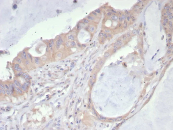
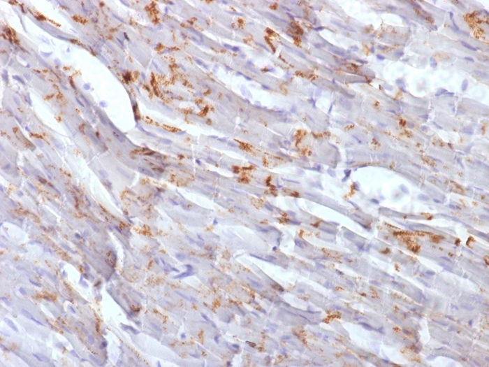
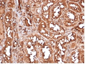
| Immunohistochemistry (IHC) | 1-2ug/ml | IHC Protocol | 30 min at RT. Staining of formalin-fixed tissues requires heating tissue sections in 10mM Tris with 1mM EDTA, pH 9.0, for 45 min at 95°C followed by cooling at RT for 20 minutes |
Platelet-derived growth factor (PDGF) is a mitogen for mesenchyme- and glia-derived cells. PDGF consists of two chains, A and B, which dimerize to form functionally distinct isoforms, PGDF-AA, PDGF-AB and PDGF-BB. These three isoforms bind with different affinities to two receptor types, PDGFR-a and -β, which are endowed with protein tyrosine kinase domains. PDGFR-a can bind to both A and B subunits of PDGF, while PDGFR-β can only bind the B subunit. Ligand binding promotes either homo- or heterodimerization of the PDGF receptors in a specific manner. PDGF-AA induces the dimerization of two a receptors, PDGF-AB induces dimerization of aa and aβ and PDGF-BB induces the formation of three types of dimers, aa, aβ and ββ. Translocation of the PDGFR-β gene with the Tel gene is linked to chronic myelomonocytic leukemia (CMML), a myelodysplastic syndrome, and demonstrates the oncogenic potential of the PDGF receptors.
Should a NeoBiotechnologies antibody not meet the experimental expectations outlined on our website or datasheet, please contact our Technical Support. We unconditionally guarantee our product.
Our team of scientists are ready to help you with expert recommendations or technical assistance related to applications or product use.
Information
© 2026 NeoBiotechnologies. All Rights Reserved.
Monday-Friday: 8:00am – 8:00pm Pacific Time