Trial Size Available
Carrier-Free Available

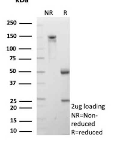
SDS-PAGE Analysis Purified CD206 Mouse Monoclonal Antibody (MRC1/9456). Confirmation of Purity and Integrity of Antibody.
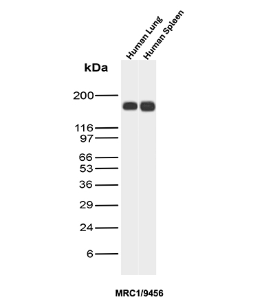
Western Blot Analysis of human lung and human spleen lysates using MRC1 Mouse Monoclonal Antibody (MRC1/9456).
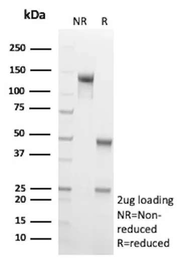
SDS-PAGE Analysis of Purified CD206 Mouse Monoclonal Antibody (MRC1/9463). Confirmation of Purity and Integrity of Antibody.

SDS-PAGE Analysis Purified CD35 Mouse Monoclonal Antibody (CR1/9444). Confirmation of Purity and Integrity of Antibody.
| Document Name | |
|---|---|
| Datasheet | Download Here |
| Material Safety Data Sheet | Download Here |
| Applications | Tested Dilutions | Protocol | Note |
|---|---|---|---|
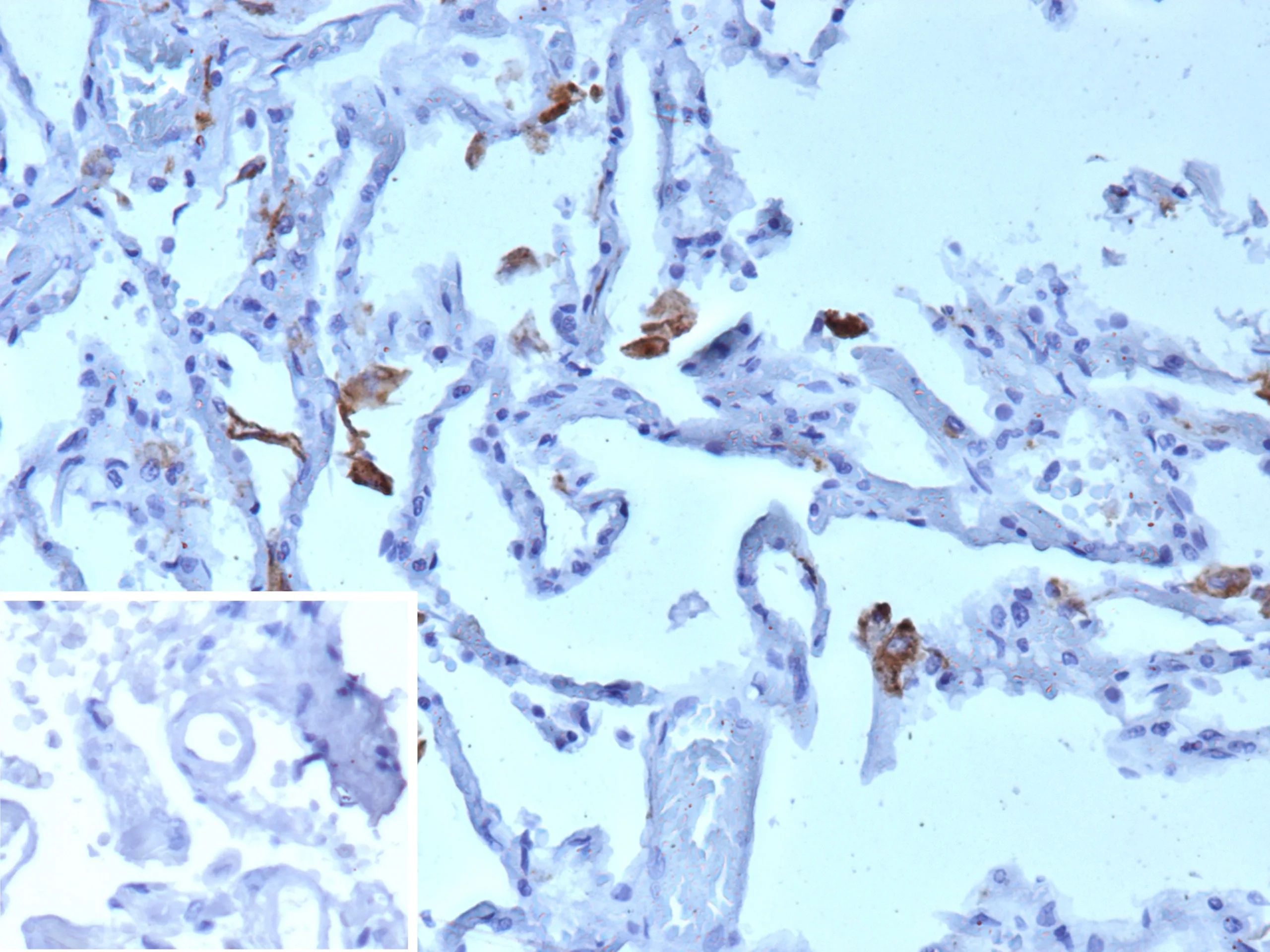
| Immunohistochemistry (IHC) | 1-2ug/ml | IHC Protocol | 30 min at RT. Staining of formalin-fixed tissues requires heating tissue sections in 10mM Tris with 1mM EDTA, pH 9.0, for 45 min at 95°C followed by cooling at RT for 20 minutes |
| Western Blot (WB) | 2-4ug/ml | Western Blot Protocol |
CD206, also known as macrophage mannose receptor type C (MMR or MRC1), is a type I membrane receptor protein. It is an phagocytic and endocytic receptor that can recognize carbohydrate ligands in target molecules. The extracellular portion of the protein includes eight C-type carbohydrate recognition domains (CRD) which are clustered together to achieve higher affinity binding to saccharides. CD206 is found on macrophages and on endothelial cells of
the liver and is the only known example of a C-type lectin that contains multiple C-type CRDs. CD206 mediates the endocytosis of glycoproteins by macrophages and binds high-mannose structures on the surface of potentially pathogenic viruses, fungi and bacteria enabling them to be neutralized by phagocytic engulfment. During inflammation, CD206 is crucial for rapid clearance of several mannose-bearing serum glycoproteins but does not regulate the initiation of inflammation. CD206 is primarily expressed in mature tissue macrophages and immature dendritic cells, as well as hepatic and lymphatic endothelial cells, retinal pigmental epithelium (RPE) and mesangial cells.
Should a NeoBiotechnologies antibody not meet the experimental expectations outlined on our website or datasheet, please contact our Technical Support. We unconditionally guarantee our product.
Our team of scientists are ready to help you with expert recommendations or technical assistance related to applications or product use.
Information
© 2026 NeoBiotechnologies. All Rights Reserved.
Monday-Friday: 8:00am – 8:00pm Pacific Time