Trial Size Available
Carrier-Free Available

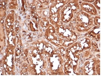
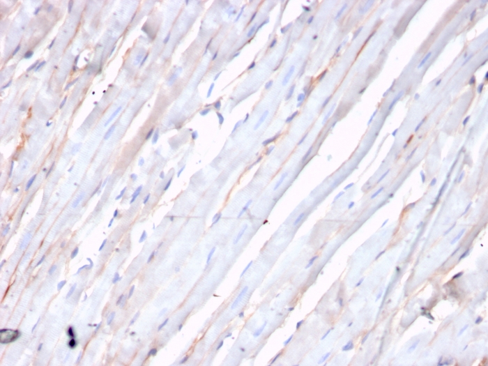
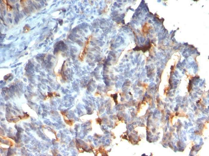
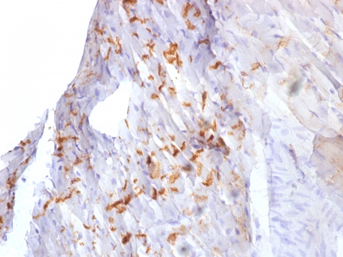
Formalin-fixed, paraffin-embedded human small intestine stained with MUC-1 Recombinant Mouse Monoclonal Antibody (rMUC1/955).
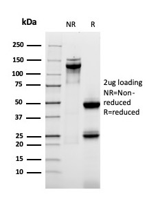
SDS-PAGE Analysis of Purified MUC-1 Recombinant Mouse Monoclonal Antibody (rMUC1/955). Confirmation of Purity and Integrity of Antibody.
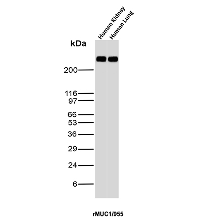
Western Blot Analysis of Human kidney and Human Lung tissue lysates using MUC-1 Recombinant Mouse Monoclonal Antibody (MUC1/955).
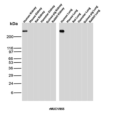
Western Blot Analysis of Human Kidney, Mouse Kidney, Rat Kidney, Hamster Kidney, Guinea pig Kidney, Rabbit Kidney, Human Lung, Mouse Lung, Rat Lung, Hamster Lung, Guinea pig Lung and Rabbit Lung tissue lysates using MUC-1 / CA15-3 / EMA Recombinant Mouse Monoclonal Antibody (MUC1/955).
| Document Name | |
|---|---|
| Datasheet | Download Here |
| Material Safety Data Sheet | Download Here |
| Applications | Tested Dilutions | Protocol | Note |
|---|---|---|---|
| Immunohistochemistry (IHC) | 1-2ug/ml | IHC Protocol | 30 min at RT. Staining of formalin-fixed tissues requires heating tissue sections in 10mM Tris with 1mM EDTA, pH 9.0, for 45 min at 95°C followed by cooling at RT for 20 minutes |
| Western Blot (WB) | 2-4ug/ml | Western Blot Protocol |
This MAb reacts with MUC1. The dominant epitope of this MAb has not yet been determined. MUC1 is a large cell surface mucin glycoprotein expressed by most glandular and ductal epithelial cells and some hematopoietic cell lineages. It is expressed on most secretory epithelium, including mammary gland and some hematopoietic cells. It is expressed abundantly in lactating mammary glands and over expressed abundantly in >90% breast carcinomas and metastases. Transgenic MUC1 has been shown to associate with all four c-erbB receptors and localize with c-erbB1 (EGFR) in lactating glands. The MUC1 gene contains seven exons and produces several different alternatively spliced variants. The major expressed form of MUC1 uses all seven exons and is a type 1 transmembrane protein with a large extracellular tandem repeat domain. The tandem repeat domain is highly O glycosylated and alterations in glycosylation have been shown in epithelial cancer cells. Antibody to EMA is useful as a pan-epithelial marker for detecting early metastatic loci of carcinoma in bone marrow or liver.
Should a NeoBiotechnologies antibody not meet the experimental expectations outlined on our website or datasheet, please contact our Technical Support. We unconditionally guarantee our product.
Our team of scientists are ready to help you with expert recommendations or technical assistance related to applications or product use.
Quick Links
Information
© 2026 NeoBiotechnologies.  All Rights Reserved.
Monday-Friday: 8:00am – 8:00pm Pacific Time