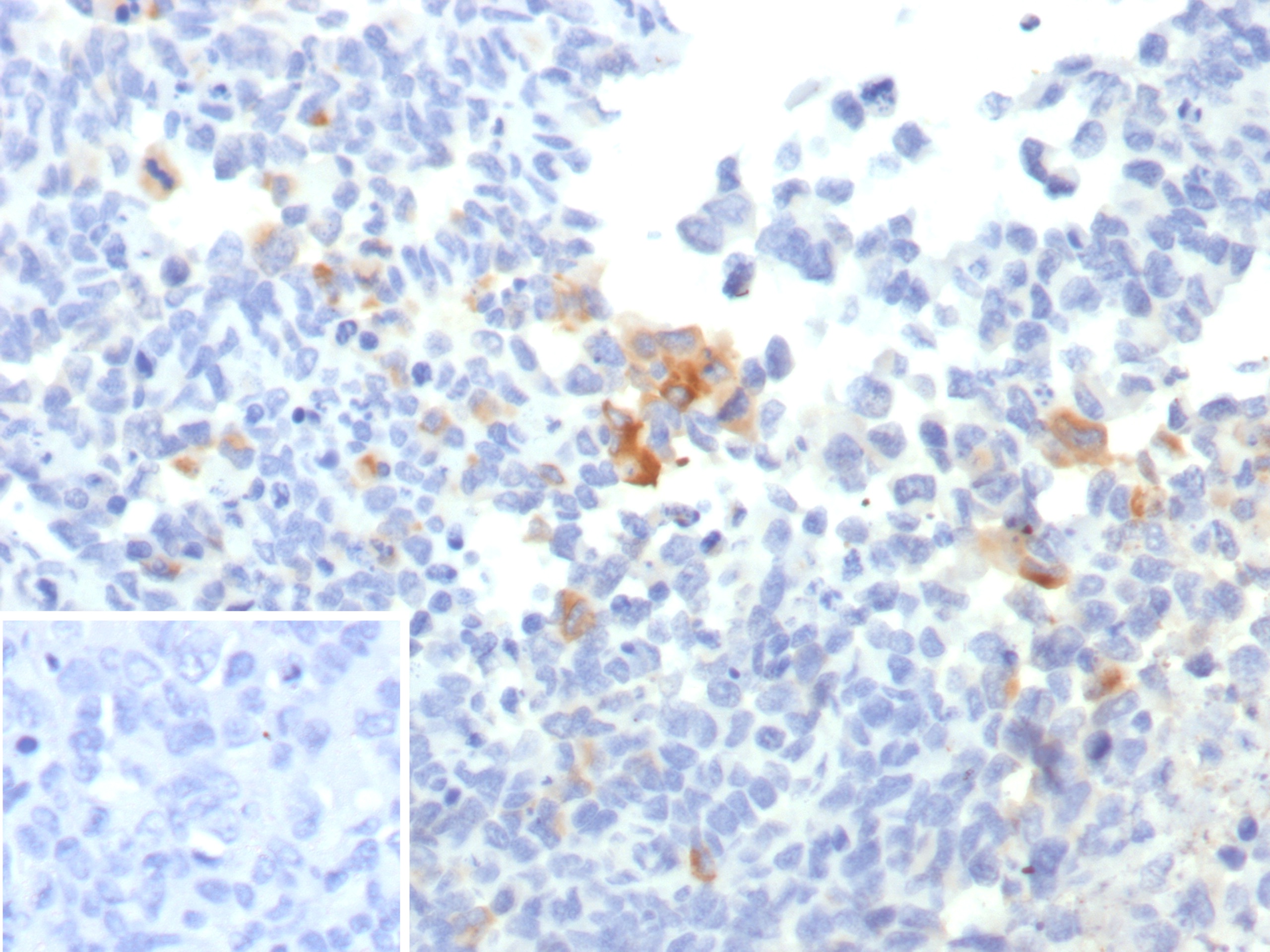
Recombinant gp100 / Melanosome / PMEL17 / SILV (Melanoma Marker) Antibody [PMEL/7554R]

Additional information

Catalog Number: 6490-RBM14
Gene:

Host:

Reactivity:

In Stock

Trial Size Available

Carrier-Free Available

In Stock
Catalog Number Formulation Size Price
6490-RBM14-P0
Purified Ab with BSA and Azide at 200ug/ml
20ug
$259.00
6490-RBM14-P1
Purified Ab with BSA and Azide at 200ug/ml
100ug
$559.00
6490-RBM14-P1ABX
Purified Ab WITHOUT BSA or Azide at 1.0mg/ml
100ug
$559.00
Flat Rate Domestic: $95 | Orders outside the US - Contact Us for Order Information | Ships next business day
  • IHC
  • WB

Documents

Document Name
Datasheet Download Here
Material Safety Data Sheet Download Here

Applications & Dilutions

Applications Tested Dilutions Protocol Note
Immunohistochemistry (IHC) 1-2ug/ml IHC Protocol 30 min at RT. Staining of formalin-fixed tissues requires heating tissue sections in 10mM Tris with 1mM EDTA, pH 9.0, for 45 min at 95°C followed by cooling at RT for 20 minutes
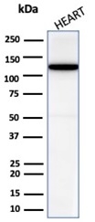
Western Blot (WB) 2-4ug/ml Western Blot Protocol

Product Properties

Host
Rabbit
Species Reactivity
Clone
PMEL/7554R
Isotype
IgG
Light Chain
Kappa
Cellular Localization
Endoplasmic reticulum membrane, Endosome, Golgi apparatus, Melanosome, Multivesicular body, Secreted, cis-Golgi network membrane, Extracellular vesicle
Gene Name
Positive Control
Skin: A moderate to strong cytoplasmic PMEL immunostaining should be seen in all melanocytes of the skin.
Immunogen
Recombinant fragment (around aa647-661) of the human PMEL17 protein (exact sequence is proprietary)
Expression System
HEK293
Purification
Protein A
Storage & Stability
Antibody with azide - store at 2 to 8 °C. Antibody without azide - store at -20 to -80 °C. Antibody is stable for 24 months. Non-hazardous. No MSDS required.
Supplied as
200ug/ml of Ab produced in a mammalian-based expression system. Prepared in 10mM PBS with 0.05% BSA & 0.05% azide. Also available WITHOUT BSA & azide at 1.0mg/ml.
Limitations
This antibody is available for research use only and is not approved for use in diagnosis.

Related Antibodies

Other Information

Functions
Warranty
There are no warranties, expressed or implied, which extend beyond this description. Company is not liable for any personal injury or economic loss resulting from this product.
Entrez Gene ID
SwissProt
Chromosome Location
12q13.2
Mol. Weight of Antigen
70.25kDa
Alternate Names
Melanocyte protein PMEL, D12S53E|HMB-45|HMB45|ME20|ME20-M|ME20M|P1|P100|PMEL17|SI|SIL|SILV|gp100

Summary

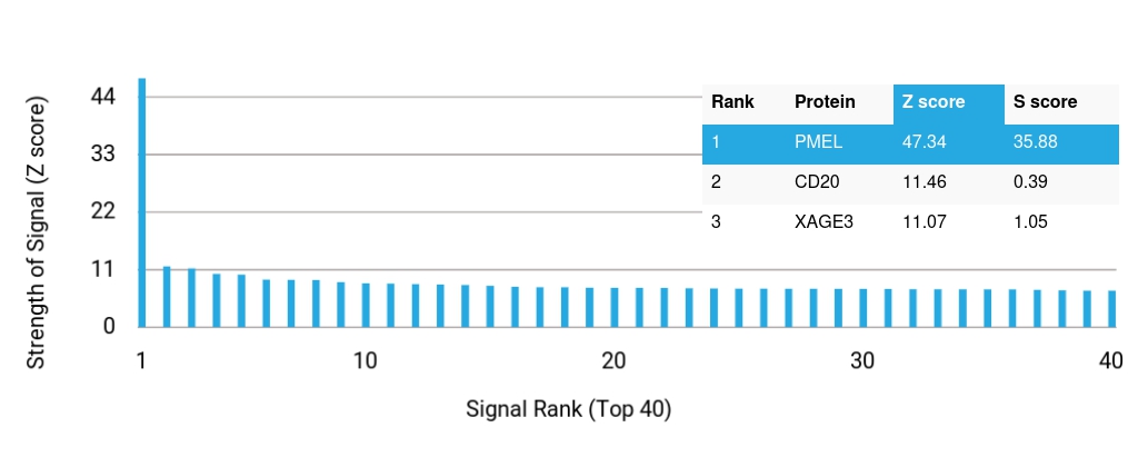
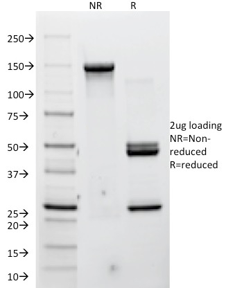
The gp100 molecule is a 100kDa glycosylated protein that is cleaved into a small (26kDa) carboxy-terminal fragment and a larger amino- terminal section (60 64 kDa), which is subsequently cleaved to generate 26kDa and 34 38kDa fragments. By immunohistochemistry, it specifically recognizes a protein in melanocytes and melanomas. This MAb reacts with junctional and blue nevus cells and variably with fetal and neonatal melanocytes. Intradermal nevi, normal adult melanocytes, and non-melanocytic cells are negative. It does not stain tumor cells of epithelial, lymphoid, glial, or mesenchymal origin. Metastatic amelanotic melanoma can often be confused with a variety of poorly differentiated carcinomas, large cell lymphomas, and sarcomas using H & E stains alone. It is also difficult to differentiate melanoma from spindle cell carcinomas and various types of mesenchymal neoplasms. This MAb stains fetal and neonatal melanocytes, junctional and blue nevus cells, and malignant melanoma. This MAb also stains Angiomyolipoma (PEComa).

Worry-Free Guarantee

Should a NeoBiotechnologies antibody not meet the experimental expectations outlined on our website or datasheet, please contact our Technical Support. We unconditionally guarantee our product.

Need Technical Help?

Our team of scientists are ready to help you with expert recommendations or technical assistance related to applications or product use.