


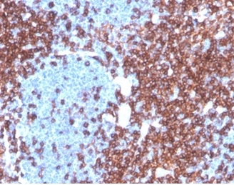
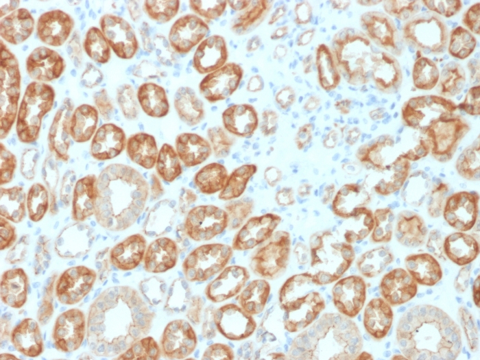
Formalin-fixed, paraffin-embedded human lymph node stained with CD4 Recombinant Rabbit Monoclonal Antibody (CD4/3619R) at 2ug/ml. HIER: Tris/EDTA, pH9.0, 45min. 2°C: HRP-polymer, 30min. DAB, 5min.

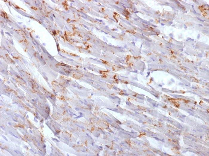
Formalin-fixed, paraffin-embedded human tonsil stained with CD4 Recombinant Rabbit Monoclonal Antibody (CD4/3619R) at 2ug/ml. HIER: Tris/EDTA, pH9.0, 45min. 2°C: HRP-polymer, 30min. DAB, 5min.
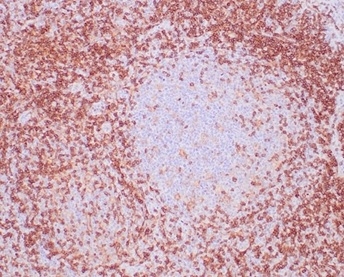
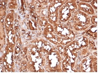
Formalin-fixed, paraffin-embedded human lymph node stained with CD4 Recombinant Rabbit Monoclonal Antibody (CD4/3619R) at 2ug/ml. HIER: Tris/EDTA, pH9.0, 45min. 2°C: HRP-polymer, 30min. DAB, 5min.
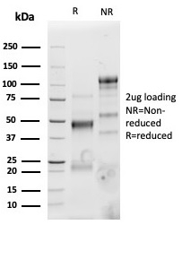
SDS-PAGE Analysis of Purified CD4 Recombinant Rabbit Monoclonal Antibody (CD4/3619R). Confirmation of Purity and Integrity of Antibody.

Analysis of Protein Array containing more than 19,000 full-length human proteins using CD4-Monospecific Recombinant Rabbit Monoclonal Antibody (CD4/3619R). Z- and S- Score: The Z-score represents the strength of a signal that a monoclonal antibody (MAb) (in combination with a fluorescently-tagged anti-IgG secondary antibody) produces when binding to a particular protein on the HuProtTM array. Z-scores are described in units of standard deviations (SD's) above the mean value of all signals generated on that array. If targets on HuProtTM are arranged in descending order of the Z-score, the S-score is the difference (also in units of SD's) between the Z-score. S-score therefore represents the relative target specificity of a MAb to its intended target. A MAb is considered to specific to its intended target, if the MAb has an S-score of at least 2.5. For example, if a MAb binds to protein X with a Z-score of 43 and to protein Y with a Z-score of 14, then the S-score for the binding of that MAb to protein X is equal to 29.

Western Blot Analysis of human Tonsil tissue lysate using CD4 Recombinant Rabbit Monoclonal Antibody (CD4/3619R).
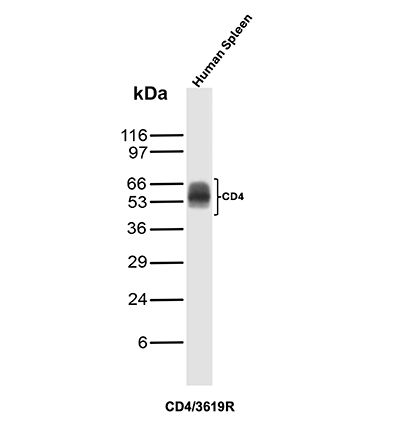
Western Blot Analysis of Human Spleen tissue lysate using CD4 Recombinant Rabbit Monoclonal Antibody (CD4/3619R).

Western Blot Analysis of Spleen tissue lysates of different species using CD4 Recombinant Rabbit Monoclonal Antibody (CD4/3619R).

 4
4
 1
1
 5
5
 5
5
 7
7
 3
3
 3
3
 3
3

Monday-Friday: 8:00am – 8:00pm Pacific Time