


Trial Size Available

Carrier-Free Available
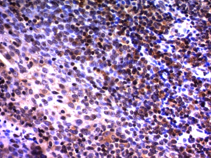
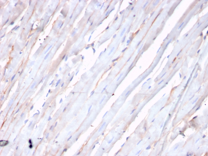
Formalin-fixed, paraffin-embedded human tonsil stained with NuMA Monoclonal Antibody (A73-B/D12)

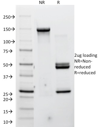
SDS-PAGE Analysis of Purified Nuclear mitotic apparatus protein 1 Mouse Monoclonal Antibody (A73-B/D12). Confirmation of Purity and Integrity of Antibody.
| Document Name | |
|---|---|
| Datasheet | Download Here |
| Material Safety Data Sheet | Download Here |
| Applications | Tested Dilutions | Protocol | Note |
|---|---|---|---|
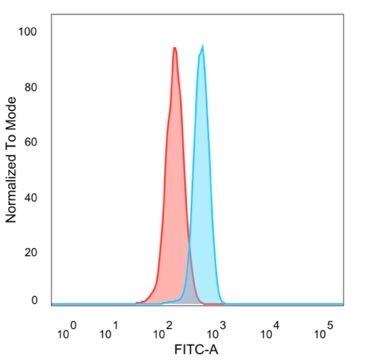
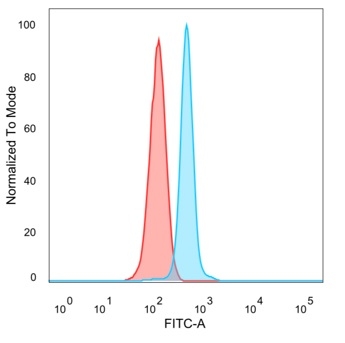
| Flow Cytometry (Flow) | 1-2ug/million cells | Flow Cytometry Protocol | |
| Immunofluorescence (IF) | 1-3ug/ml | ||
| Immunohistochemistry (IHC) | 1-2ug/ml | IHC Protocol | 30 min at RT. Staining of formalin-fixed tissues requires heating tissue sections in 10mM Tris with 1mM EDTA, pH 9.0, for 45 min at 95°C followed by cooling at RT for 20 minutes |
 0
0

 4
4
 5
5
 2
2
 3
3
 3
3
 3
3
 3
3
Recognizes a phosphorylated protein of 228kDa, identified as nuclear mitotic apparatus protein (NuMA). Its epitope is resistant to phosphatases. NuMA is intra-nuclear protein and present in nucleus during interphase. At the onset of mitosis, it redistributes from the nucleus to two centrosomal structures that later will become part of the mitotic spindle pole. After anaphase, the protein redistributes from the spindle polar region into reforming nucleus. NuMA is an essential protein during mitosis for the terminal phases of chromosome separation and/or nuclear reassembly. Recently a study shows that NuMA is cleaved to a 180 to 200kDa during apoptosis. Chromosomal translocation of this gene with the RARA (retinoic acid receptor, alpha) gene on chromosome 17 has been detected in patients with acute promyelocytic leukemia.
Should a NeoBiotechnologies antibody not meet the experimental expectations outlined on our website or datasheet, please contact our Technical Support. We unconditionally guarantee our product.
Our team of scientists are ready to help you with expert recommendations or technical assistance related to applications or product use.

Quick Links
Information
© 2026 NeoBiotechnologies. All Rights Reserved.
Monday-Friday: 8:00am – 8:00pm Pacific Time