Trial Size Available
Carrier-Free Available

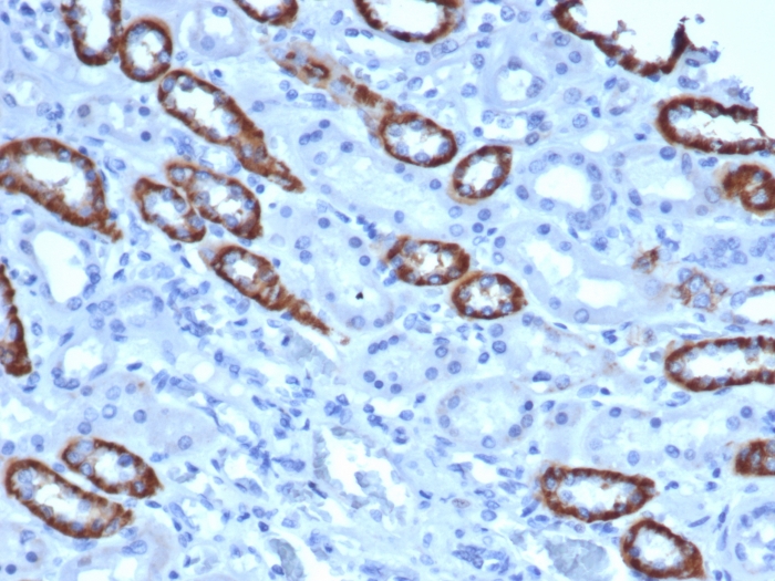
Formalin-fixed, paraffin-embedded human Liver stained with Cytochrome C Recombinant Rabbit Monoclonal Antibody (CYCS/3128R).

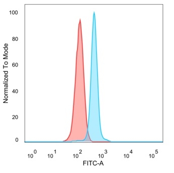
Flow Cytometric Analysis of PFA fixed HeLa cells using Cytochrome C Recombinant Rabbit Monoclonal Antibody (CYCS/3128R) followed by Goat anti-rabbit IgG-CF488 (Blue); Goat anti-mouse IgG-CF488 is Control (Red).

Western Blot Analysis of Heart tissue lysate using Cytochrome C Recombinant Rabbit Monoclonal Antibody (CYCS/3128R).

Immunofluorescence Analysis of PFA fixed HeLa cells labeling Cytochrome C Recombinant Rabbit Monoclonal Antibody (CYCS/3128R) followed by Goat anti-rabbit IgG-CF488 (Green)

Western Blot Analysis of Human Intestine, Human Heart, Human Skeletal Muscle, A549, HeLa and Jurkat lysates using Cytochrome C Recombinant Rabbit Monoclonal Antibody (CYCS/3128R).
| Document Name | |
|---|---|
| Datasheet | Download Here |
| Material Safety Data Sheet | Download Here |
| Applications | Tested Dilutions | Protocol | Note |
|---|---|---|---|
| Immunofluorescence (IF) | 1-3ug/ml | ||
| Immunohistochemistry (IHC) | 1-2ug/ml | IHC Protocol | 30 min at RT. Staining of formalin-fixed tissues requires heating tissue sections in 10mM Tris with 1mM EDTA, pH 9.0, for 45 min at 95°C followed by cooling at RT for 20 minutes |
| Western Blot (WB) | 2-4ug/ml | Western Blot Protocol |
It recognizes an epitope within amino acids 93-104 of pigeon Cytochrome C, a well-characterized mobile electron transport protein that is essential to energy conversion in all aerobic organisms. In mammalian cells, this highly conserved protein is normally localized to the mitochondrial inter-membrane space. More recent studies have identified cytosolic cytochrome c as a factor necessary for activation of apoptosis. During apoptosis, cytochrome c is trans-located from the mitochondrial membrane to the cytosol, where it is required for activation of caspase-3 (CPP32). Overexpression of Bcl-2 has been shown to prevent the translocation of cytochrome c, thereby blocking the apoptotic process. Overexpression of Bax has been shown to induce the release of cytochrome c and to induce cell death. The release of cytochrome c from the mitochondria is thought to trigger an apoptotic cascade, whereby Apaf-1 binds to Apaf-3 (caspase-9) in a cytochrome c-dependent manner, leading to caspase-9 cleavage of caspase-3. This MAb recognizes total cytochrome C which includes both apocytochrome (i.e. cytochrome in the cytosol without heme attached) and holocytochrome (i.e. cytochrome in the mitochondria with heme attached).
Should a NeoBiotechnologies antibody not meet the experimental expectations outlined on our website or datasheet, please contact our Technical Support. We unconditionally guarantee our product.
Our team of scientists are ready to help you with expert recommendations or technical assistance related to applications or product use.
Information
© 2026 NeoBiotechnologies. All Rights Reserved.
Monday-Friday: 8:00am – 8:00pm Pacific Time