Trial Size Available
Carrier-Free Available

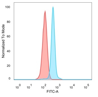
Flow cytometric analysis of cell-surface Nectin-4 on T47D (human ductal breast epithelial tumor epithelial) cells. T47D cells were stained with recombinant rabbit anti–Nectin-4 monoclonal antibody (NECTIN4/13438R) at dilutions of 1:50 (4 μg/ml; dark red), 1:100 (2 μg/ml; orange), 1:200 (1 μg/ml; light green) and 1:400 (0.5 μg/ml; dark green), then incubated with goat anti-rabbit IgG–CF488. CF488 fluorescence intensity (log₁₀ scale; x-axis) is plotted against relative cell count (y-axis). Progressive rightward shifts with increasing antibody concentration demonstrate dilution-dependent binding of Nectin-4. The blue histogram corresponds to the isotype control.
| Document Name | |
|---|---|
| Datasheet | Download Here |
| Material Safety Data Sheet | Download Here |
| Applications | Tested Dilutions | Protocol | Note |
|---|---|---|---|
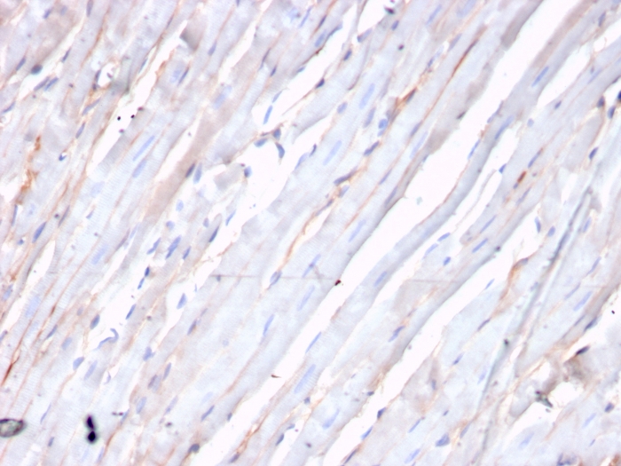
| Immunohistochemistry (IHC) | 1-2ug/ml | IHC Protocol | 30 min at RT. Staining of formalin-fixed tissues requires heating tissue sections in 10mM Tris with 1mM EDTA, pH 9.0, for 45 min at 95°C followed by cooling at RT for 20 minutes |
| Western Blot (WB) | 2-4ug/ml | Western Blot Protocol |
Homologous to the poliovirus receptor (PVR/CD155), the nectin immunoglobulin superfamily comprises four known isoforms (-1, -2, -3, and -4). The ectodomain of nectin family members comprises three Ig-like domains (V, C, C). Nectins localize at the adherens junctions (AJ) in epithelial and endothelial cells where they serve as adhesion molecules. Actin-based AJs play a role in mechanical adhesion, cellular morphogenesis and cellular differentiation. Nectin associates with the Actin cytoskeleton through its interaction with the Actin filament-binding protein afadin. Nectin 4 and afadin co-localize at cadherin-based adherens junctions in MDCKII epithelial cells. Nectin 4 and Nectin 3 share a common binding region in the V domain of Nectin 1 and thus compete for Nectin 1 binding. The Nectin 3/4 binding domain maps to the C-C’-C”-D b strands of the V domain of Nectin 1. Unlike other nectins, which are more widely expressed, Nectin 4 is mainly expressed in the placenta.
Should a NeoBiotechnologies antibody not meet the experimental expectations outlined on our website or datasheet, please contact our Technical Support. We unconditionally guarantee our product.
Our team of scientists are ready to help you with expert recommendations or technical assistance related to applications or product use.
Information
© 2026 NeoBiotechnologies. All Rights Reserved.
Monday-Friday: 8:00am – 8:00pm Pacific Time