| Catalog Number: | 4654-MSM1X |
|---|---|
| Reactivity: | |
| Gene: | |
| Host: |
Trial Size Available
Carrier-Free Available
| Document Name | |
|---|---|
| Datasheet | Download Here |
| Material Safety Data Sheet | Download Here |
| Applications | Tested Dilutions | Protocol | Note |
|---|---|---|---|
| Flow Cytometry (Flow) | 1-2ug/million cells | Flow Cytometry Protocol | |
| Immunofluorescence (IF) | 1-3ug/ml | ||
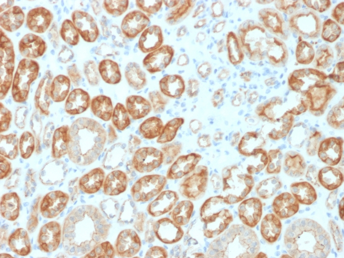
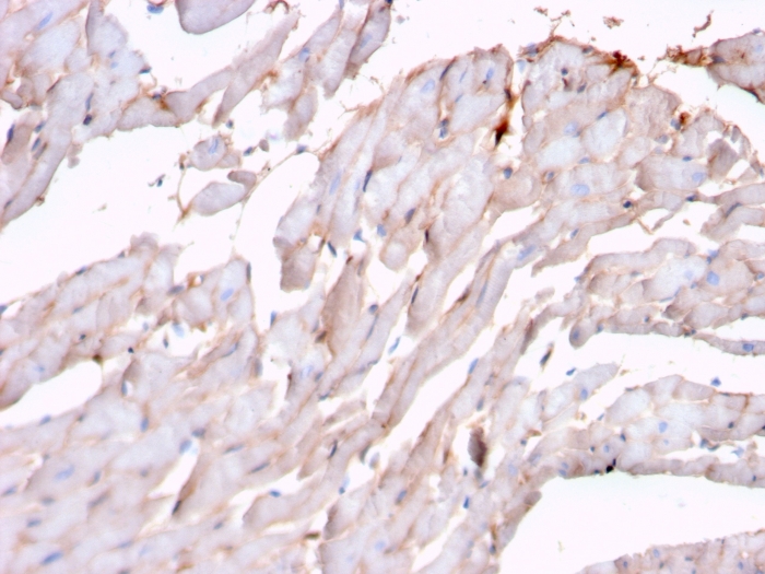
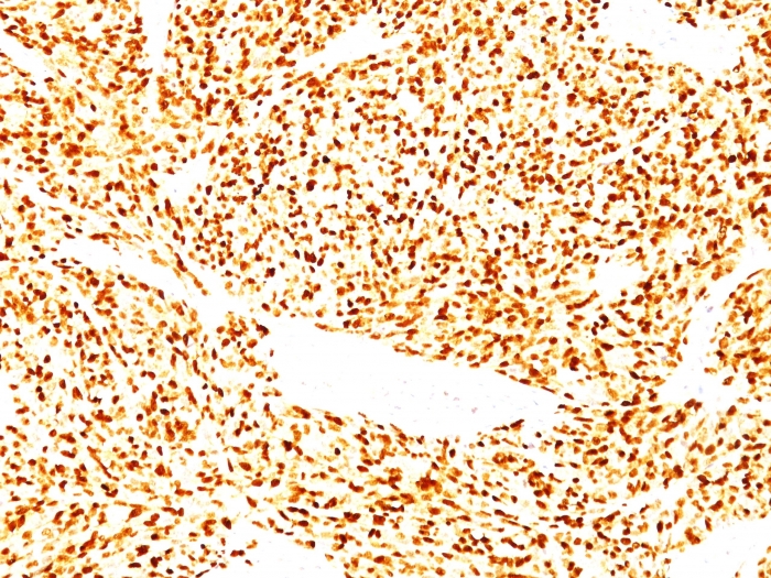
| Immunohistochemistry (IHC) | 1-2ug/ml | IHC Protocol | 30 min at RT. Staining of formalin-fixed tissues requires heating tissue sections in 10mM Tris with 1mM EDTA, pH 9.0, for 45 min at 95°C followed by cooling at RT for 20 minutes |
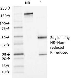
Recognizes a phosphor-protein of 45kDa, identified as MyoD1. The epitope of this MAb maps between amino acid 180-189 in the C-terminal of Mouse MyoD1 protein. It does not cross react with myogenin, Myf5, or Myf6. Antibody to MyoD1 labels the nuclei of myoblasts in developing muscle tissues. MyoD1 is not detected in normal adult tissue, but is highly expressed in the tumor cell nuclei of rhabdomyosarcomas. Occasionally nuclear expression of MyoD1 is seen in ectomesenchymoma and a subset of Wilm’s tumors. Weak cytoplasmic staining is observed in several non-muscle tissues, including glandular epithelium and also in rhabdomyosarcomas, neuroblastomas, Ewing’s sarcomas and alveolar soft part sarcomas.
Should a NeoBiotechnologies antibody not meet the experimental expectations outlined on our website or datasheet, please contact our Technical Support. We unconditionally guarantee our product.
Our team of scientists are ready to help you with expert recommendations or technical assistance related to applications or product use.
Information
© 2026 NeoBiotechnologies. All Rights Reserved.
Monday-Friday: 8:00am – 8:00pm Pacific Time