Trial Size Available
Carrier-Free Available

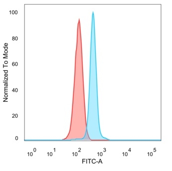
Flow cytometric analysis of PFA-fixed HeLa cells. MEF2D Mouse Monoclonal Antibody (PCRP-MEF2D-3A4) followed by goat anti-mouse IgG-CF488 (blue); isotype control (red).

Immunofluorescence Analysis of PFA-fixed HeLa cells stained using MEF2D Mouse Monoclonal Antibody (PCRP-MEF2D-3A4) followed by goat anti-mouse IgG-CF488 (green). CF640A phalloidin (red).

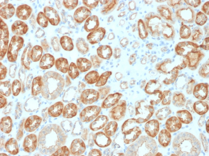
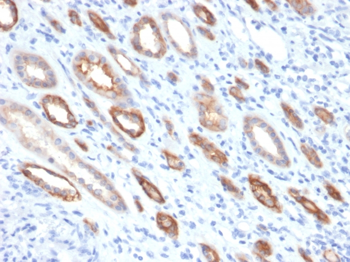
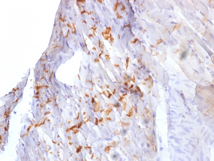
Formalin-fixed, paraffin-embedded human rhabdomyosarcoma stained with MEF2D Mouse Monoclonal Antibody (PCRP-MEF2D-3A4). HIER: Tris/EDTA, pH9.0, 45min. 2 °: HRP-polymer, 30min. DAB, 5min.

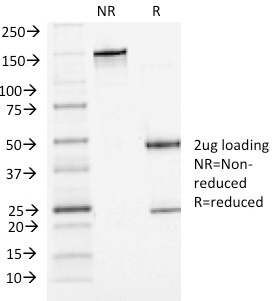
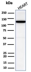
SDS-PAGE Analysis MEF2D Mouse Monoclonal Antibody (PCRP-MEF2D-3A4). Confirmation of Purity and Integrity of Antibody.
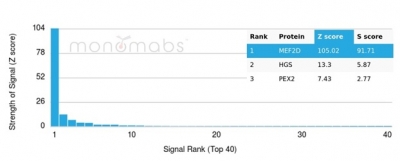
Analysis of Protein Array containing more than 19,000 full-length human proteins using MEF2D-Monospecific Mouse Monoclonal Antibody (PCRP-MEF2D-3A4). Z- and S- Score: The Z-score represents the strength of a signal that a monoclonal antibody (MAb) (in combination with a fluorescently-tagged anti-IgG secondary antibody) produces when binding to a particular protein on the HuProtTM array. Z-scores are described in units of standard deviations (SD's) above the mean value of all signals generated on that array. If targets on HuProtTM are arranged in descending order of the Z-score, the S-score is the difference (also in units of SD's) between the Z-score. S-score therefore represents the relative target specificity of a MAb to its intended target. A MAb is considered to specific to its intended target, if the MAb has an S-score of at least 2.5. For example, if a MAb binds to protein X with a Z-score of 43 and to protein Y with a Z-score of 14, then the S-score for the binding of that MAb to protein X is equal to 29.
| Document Name | |
|---|---|
| Datasheet | Download Here |
| Material Safety Data Sheet | Download Here |
| Applications | Tested Dilutions | Protocol | Note |
|---|---|---|---|
| Flow Cytometry (Flow) | 1-2ug/million cells | Flow Cytometry Protocol | |
| Immunofluorescence (IF) | 1-3ug/ml | ||
| Immunohistochemistry (IHC) | 1-2ug/ml | IHC Protocol | 30 min at RT. Staining of formalin-fixed tissues requires heating tissue sections in 10mM Tris with 1mM EDTA, pH 9.0, for 45 min at 95°C followed by cooling at RT for 20 minutes |
The myocyte enhancer factor-2 (MEF-2) family of transcription factors associate with co-repessors or co-activators to regulate development and function of T cells, neuronal cells and muscle cells. Four family members arise from alternatively spliced transcripts, termed MEF-2A, -2B, -2C and -2D. These members bind as homo- and heterodimers to the MEF-2 site in the promoter region of affected genes. Differential regulation in the expression of the four transcripts implies functional distinction for each duing embryogenesis and development. The process of differentiation from mesodermal precursor cells to myoblasts has led to the discovery of a variety of tissue-specific factors that regulate muscle gene expression. The myogenic basic helix-loop-helix proteins, including MyoD, myogenin, Myf-5 and MRF4, are one class of identified factors. A second family of DNA-binding regulatory proteins is the myocyte-specific enhancer factor-2 (MEF-2) family. Each of these proteins binds to the MEF-2 target DNA sequence present in the regulatory regions of many muscle-specific genes.
Should a NeoBiotechnologies antibody not meet the experimental expectations outlined on our website or datasheet, please contact our Technical Support. We unconditionally guarantee our product.
Our team of scientists are ready to help you with expert recommendations or technical assistance related to applications or product use.
Information
© 2026 NeoBiotechnologies.  All Rights Reserved.
Monday-Friday: 8:00am – 8:00pm Pacific Time