| Catalog Number: | 59-MSM1 |
|---|---|
| Reactivity: | Baboon, Cat, Chicken, Cow, Dog, Goat, Guinea Pig, Human, Monkey, Mouse, Pig, Rabbit, Rat, Sheep |
| Gene: | |
| Host: |
Trial Size Available
Carrier-Free Available

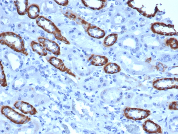
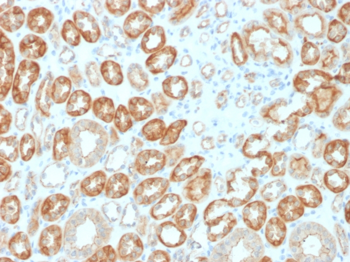
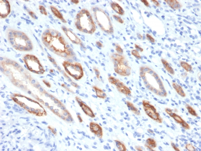
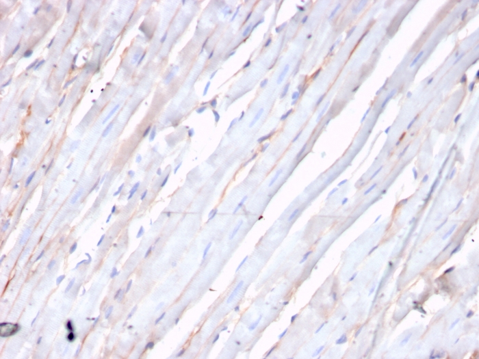
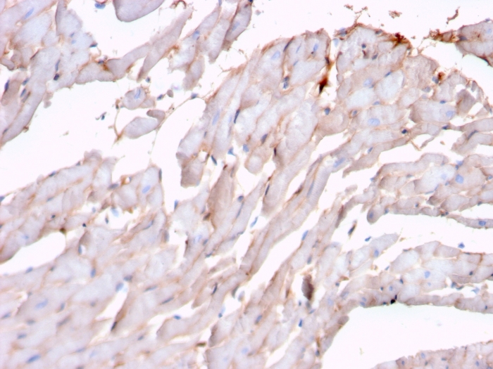
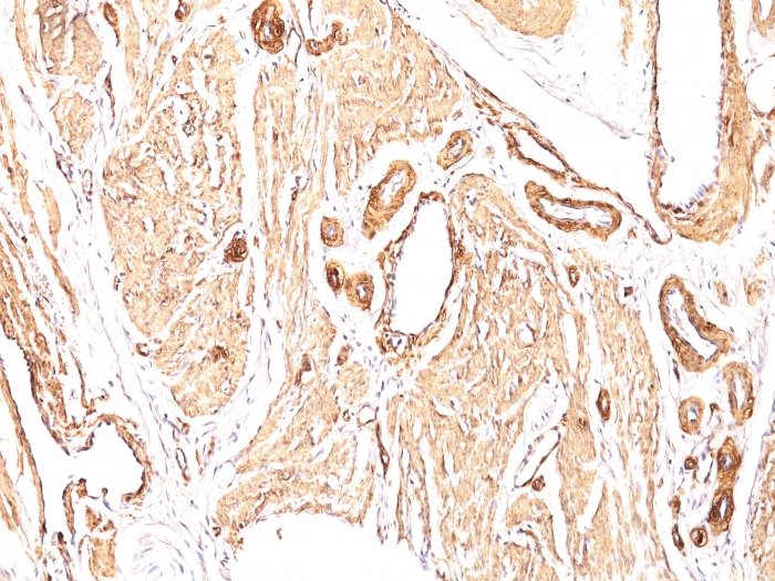
Formalin-fixed, paraffin-embedded human Leiomyosarcoma stained with Smooth Muscle Actin Mouse Monoclonal Antibody (1A4).
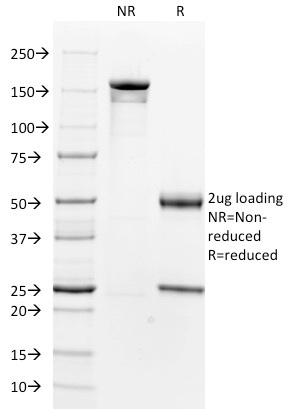
SDS-PAGE Analysis of Purified Smooth Muscle Actin Mouse Monoclonal Antibody (1A4). Confirmation of Integrity and Purity of Antibody.
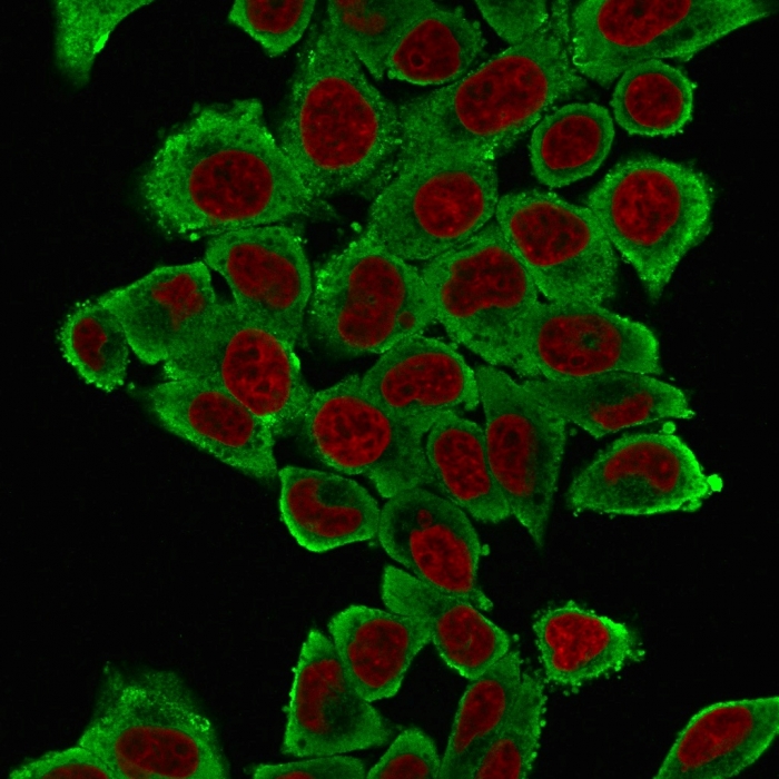
Immunofluorescence Analysis of HeLa cells labeling Smooth Muscle Actin with Smooth Muscle Actin Mouse Monoclonal Antibody (1A4) followed by goat anti-Mouse IgG-CF488 (green). The nuclear counterstain is NucSpot (red).
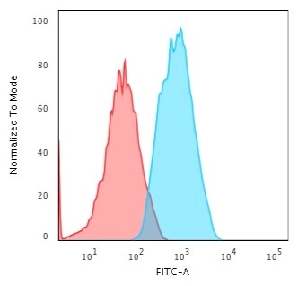
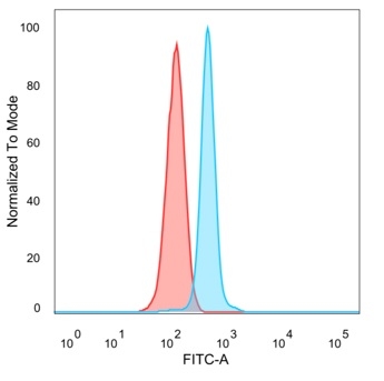
Flow Cytometric Analysis of Human HeLa cells using Smooth Muscle Actin Mouse Monoclonal Antibody (1A4) followed by Goat anti-Mouse IgG-CF488 (Blue); Isotype control (Red).
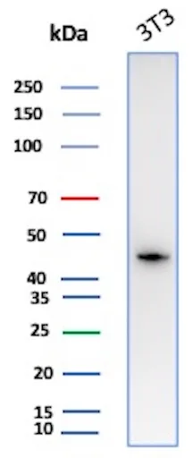
Western Blot Analysis of Mouse 3T3 lysate using ACTA2 Mouse Monoclonal Antibody (1A4)
| Document Name | |
|---|---|
| Datasheet | Download Here |
| Material Safety Data Sheet | Download Here |
| Applications | Tested Dilutions | Protocol | Note |
|---|---|---|---|
| Flow Cytometry (Flow) | 1-2ug/million cells | Flow Cytometry Protocol | |
| Immunofluorescence (IF) | 1-3ug/ml | ||
| Immunohistochemistry (IHC) | 1-2ug/ml | IHC Protocol | 30 min at RT. Staining of formalin-fixed tissues requires heating tissue sections in 10mM Tris with 1mM EDTA, pH 9.0, for 45 min at 95°C followed by cooling at RT for 20 minutes |
| Western Blot (WB) | 2-4ug/ml | Western Blot Protocol |
Actin is a major component of the cytoskeleton and is present in most cell types. This MAb is highly specific to actin from smooth muscles. Its epitope lies in the first four N-terminal amino acids. This MAb does not stain cardiac or skeletal muscle; however, it does stain myofibroblasts and myoepithelial cells. This antibody could be used together with anti-muscle specific actin and myogenin in making a diagnosis of smooth muscle and skeletal muscle tumors. In most cases of rhabdomyosarcoma, this antibody yields negative results whereas anti-muscle specific actin and myogenin are positive. Leiomyosarcomas are positive only with anti-muscle specific actin and anti-smooth muscle actin and are negative with anti-myogenin.
Should a NeoBiotechnologies antibody not meet the experimental expectations outlined on our website or datasheet, please contact our Technical Support. We unconditionally guarantee our product.
Our team of scientists are ready to help you with expert recommendations or technical assistance related to applications or product use.
Information
© 2026 NeoBiotechnologies.  All Rights Reserved.
Monday-Friday: 8:00am – 8:00pm Pacific Time