Trial Size Available
Carrier-Free Available

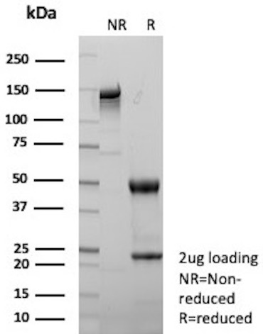
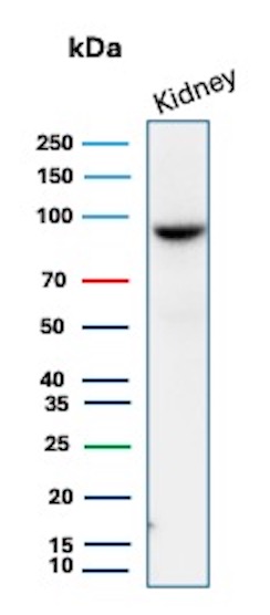
Western blot analysis of human kidney tissue lysate using STAT6 Rabbit Recombinant Monoclonal Antibody (STAT6/12798R).

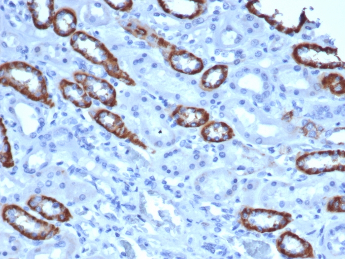
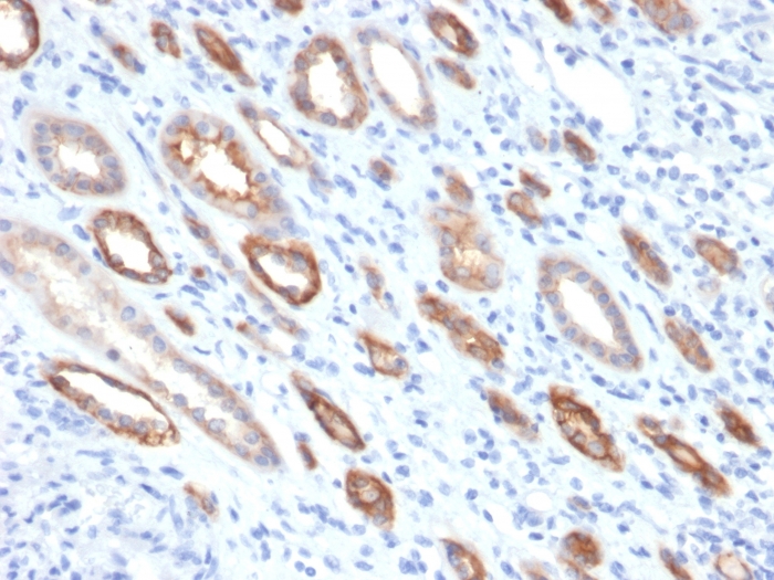
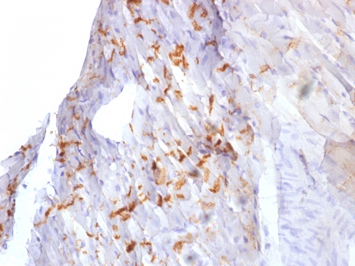
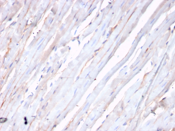
Formalin-fixed, paraffin-embedded human liposarcoma stained with STAT6 Rabbit Recombinant Monoclonal Antibody (STAT6/12798R). HIER: Tris/EDTA, pH9.0, 45min. 2°: HRP-polymer, 30min. DAB, 5min.
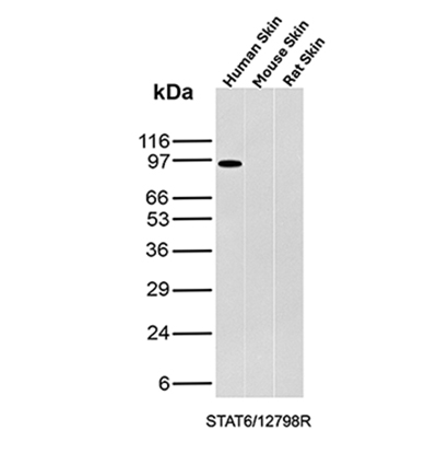
Western blot analysis of Human Skin, Mouse Skin and Rat Skin tissue lysates using STAT6 Rabbit Recombinant Monoclonal Antibody (STAT6/12798R).
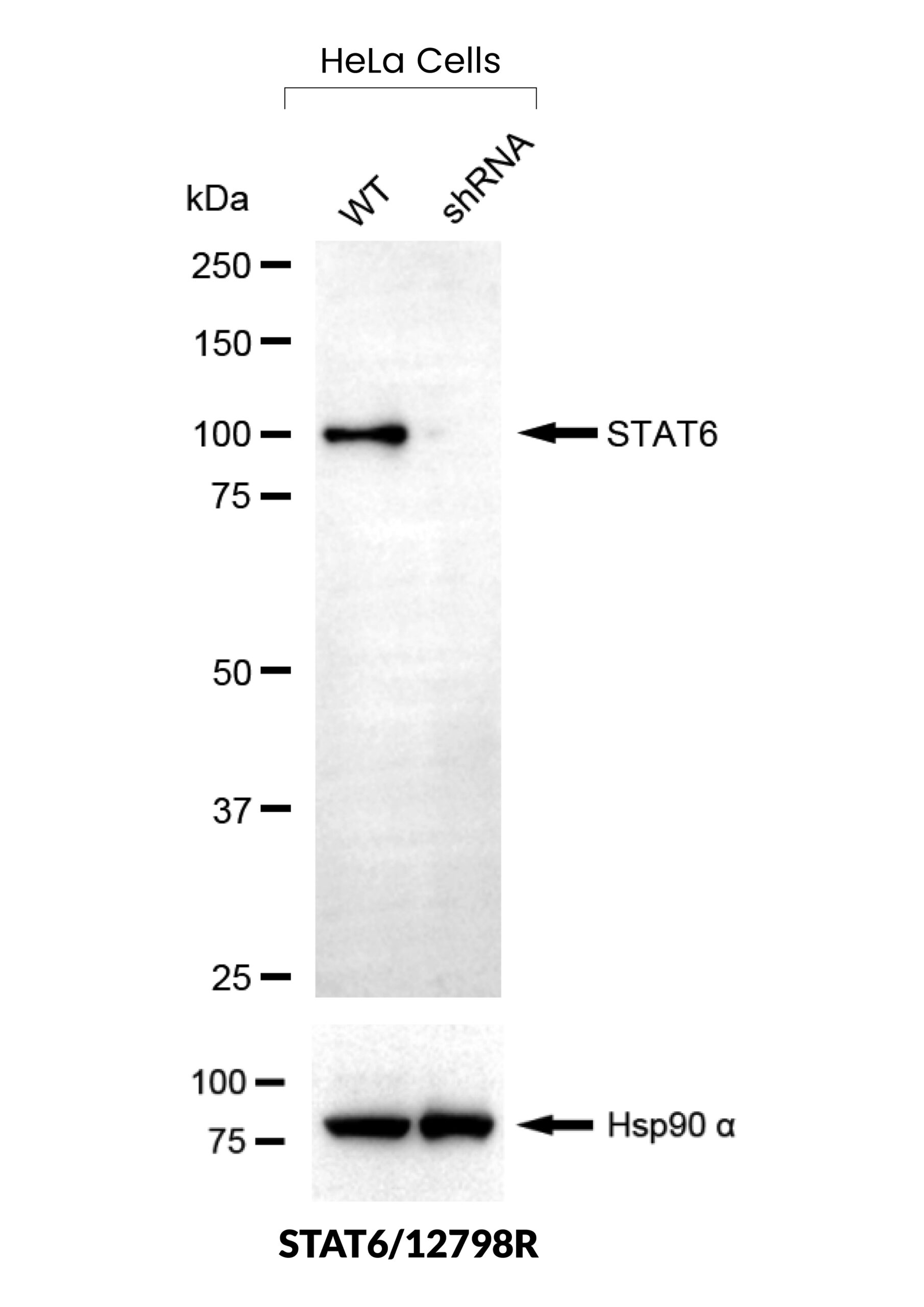
Western Blot validation of anti-STAT6 antibody. STAT6 protein expression was compared in wild-type (WT) and STAT6 shRNA Knockdown (KD) HeLa cells (30ug of total protein). Loading control is shown below.
| Document Name | |
|---|---|
| Datasheet | Download Here |
| Material Safety Data Sheet | Download Here |
| Applications | Tested Dilutions | Protocol | Note |
|---|---|---|---|
| Immunohistochemistry (IHC) | 1-2ug/ml | IHC Protocol | 30 min at RT. Staining of formalin-fixed tissues requires heating tissue sections in 10mM Tris with 1mM EDTA, pH 9.0, for 45 min at 95°C followed by cooling at RT for 20 minutes |
| Western Blot (WB) | 2-4ug/ml | Western Blot Protocol |
STAT6 is a transcription factor in the Jak/STAT signal transduction pathway responsible for mediating IL-4 immune signaling. STAT6 was recently suggested to be a reliable marker to distinguish solitary fibrous tumors from other soft tissue neoplasms. Gene fusions are common in solitary fibrous tumors. Recent next generation sequencing studies demonstrated the presence of a NAB2-STAT6 fusion, formed by an intrachromosomal inversion fusing two neighboring genes on chromosome 12q13, in 55-100% of solitary fibrous tumors, regardless of tumor morphology or anatomical site. By immunohistochemistry, nuclear STAT6 expression can discriminate solitary fibrous tumors from its morphological mimics in the meninges, including meningioma, glioblastoma, gliosarcoma, haemangioblastoma, schwannoma and haemangioma. A recent study by Cheah, et al. using the rabbit monoclonal STAT6 antibody (Clone YE361) observed expression in all solitary fibrous tumors (54/54) tested, regardless of histology, anatomical site or CD34 status. Morphological mimics of solitary fibrous tumors were negative, demonstrating 100% specificity.
Should a NeoBiotechnologies antibody not meet the experimental expectations outlined on our website or datasheet, please contact our Technical Support. We unconditionally guarantee our product.
Our team of scientists are ready to help you with expert recommendations or technical assistance related to applications or product use.
Information
© 2026 NeoBiotechnologies. All Rights Reserved.
Monday-Friday: 8:00am – 8:00pm Pacific Time