Trial Size Available
Carrier-Free Available

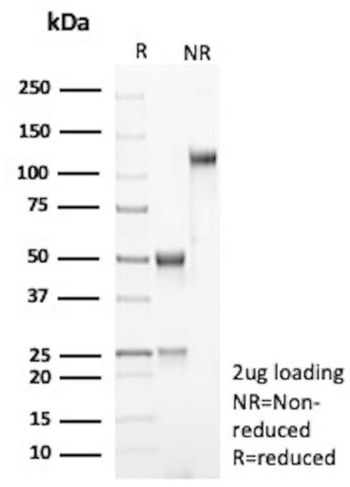
SDS-PAGE Analysis Purified PDL2 Mouse Monoclonal Antibody (PDL2/2681). Confirmation of Purity and Integrity of Antibody.
| Document Name | |
|---|---|
| Datasheet | Download Here |
| Material Safety Data Sheet | Download Here |
| Applications | Tested Dilutions | Protocol | Note |
|---|---|---|---|
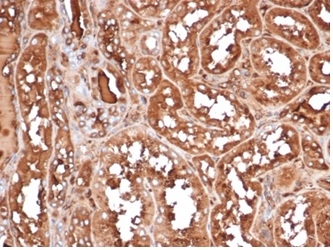
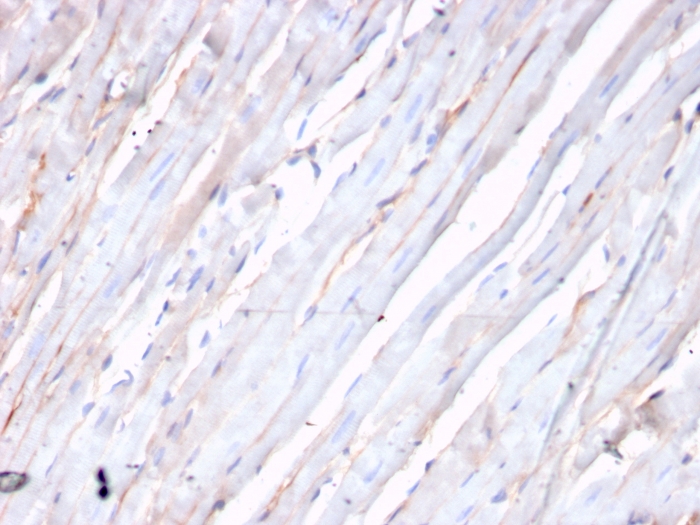
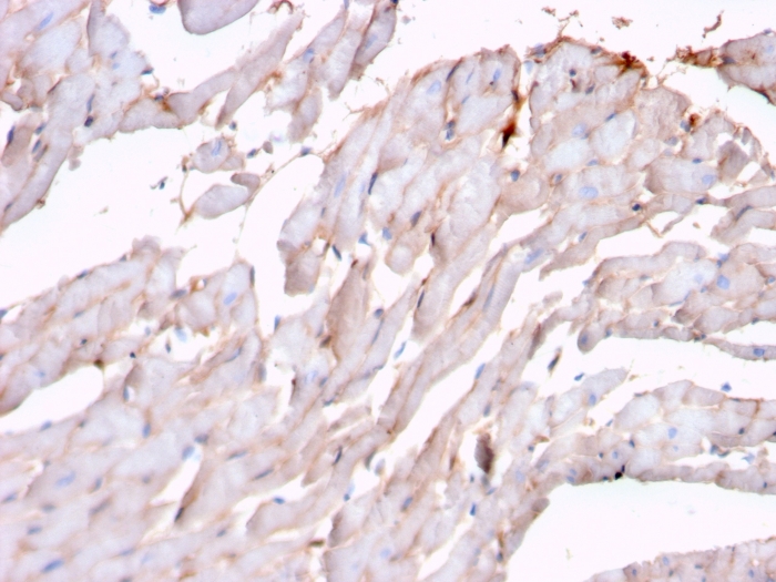
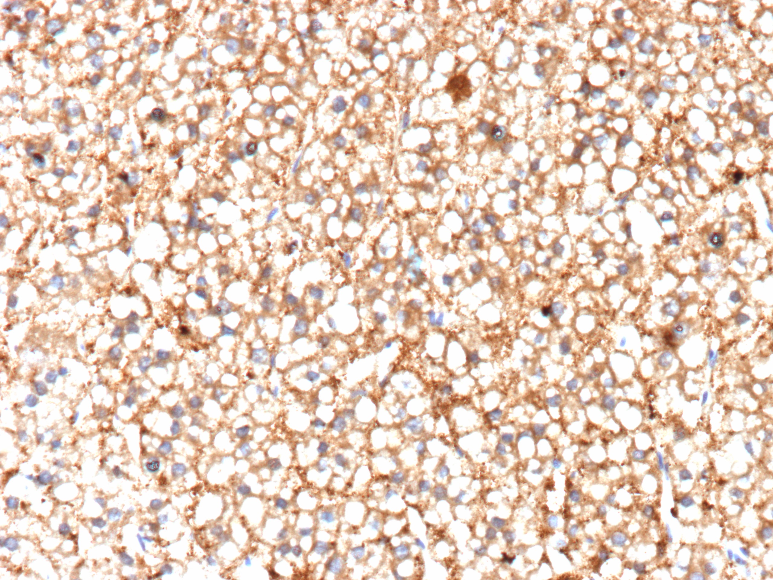
| Immunohistochemistry (IHC) | 1-2ug/ml | IHC Protocol | 30 min at RT. Staining of formalin-fixed tissues requires heating tissue sections in 10mM Tris with 1mM EDTA, pH 9.0, for 45 min at 95°C followed by cooling at RT for 20 minutes |
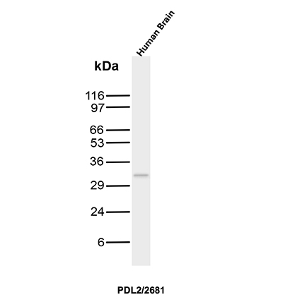
| Western Blot (WB) | 2-4ug/ml | Western Blot Protocol |
Recognizes a protein of about 31kDa, which is identified as PD-L2 (same as PDCD1LG2). Engagement of CD28 by B7-1 (CD80) or B7-2 (CD86) in the presence of antigen promotes T cell proliferation, cytokine production, differentiation of effector T cells and the induction of Bcl-x, a promoter of T cell survival. Conversely, engagement of CTLA4 by B7-1 or B7-2 may inhibit proliferation and IL-2 production. PD-L2 does not bind CD28, cytotoxic T lymphocyte A4 or ICOS (inducible co-stimulator). The constitutive expression of PD-L1 and PD-L2 on parenchymal cells of heart, lung and kidney suggests that the Pdcd-1-Pdcd-L system could provide unique negative signaling to help prevent autoimmune disease.
Should a NeoBiotechnologies antibody not meet the experimental expectations outlined on our website or datasheet, please contact our Technical Support. We unconditionally guarantee our product.
Our team of scientists are ready to help you with expert recommendations or technical assistance related to applications or product use.
Information
© 2026 NeoBiotechnologies.  All Rights Reserved.
Monday-Friday: 8:00am – 8:00pm Pacific Time